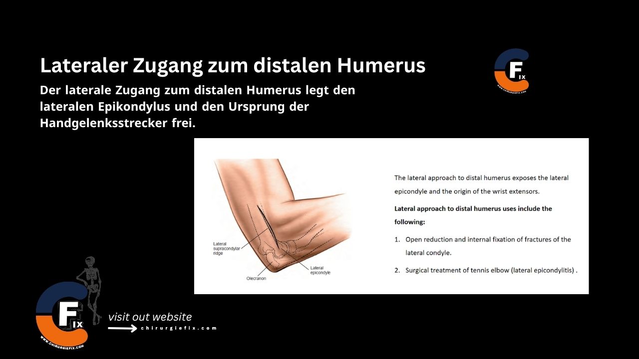
The lateral approach to the distal humerus provides exposure of the lateral epicondyle and the origin of the wrist extensors.
Indications for this approach include:
The patient is placed in the supine position on the operating table with the arm resting across the chest.
Landmarks:
Incision:
Make a curved or straight incision, 4 to 6 cm long, on the lateral side of the elbow over the lateral supracondylar ridge.
There is no true internervous plane for the lateral approach to the distal humerus. Dissection is performed between the triceps muscle and the brachioradialis muscle, both of which are innervated by the radial nerve.
Incise the deep fascia along the skin incision. Identify the plane between the brachioradialis muscle and the triceps muscle. Dissect between these two muscles down to the bone, then retract the triceps posteriorly and the brachioradialis anteriorly.
The origin of the common extensor muscle can be elevated from the lateral humerus, and the triceps can be retracted posteriorly in a similar fashion.
Proximal extension is not possible because the radial nerve crosses the planned line of dissection.
The lateral approach can be extended onto the radial head only through the intramuscular plane between the anconeus muscle (supplied by the radial nerve) and the extensor carpi ulnaris muscle (supplied by the posterior interosseous nerve). The lateral approach to the distal humerus cannot be extended further distally due to the presence of the posterior interosseous nerve, which winds around the neck of the proximal radius.
During proximal extension of the lateral approach to the distal humerus, the radial nerve is at risk as it penetrates the lateral intermuscular septum in the distal third of the arm.
The radial nerve is the continuation of the posterior cord of the brachial plexus. It begins behind the axillary artery at the shoulder, runs along the posterior wall of the axilla (on the subscapularis, latissimus dorsi, and teres major muscles), and then passes through the triangular space between the long head of the triceps and the humeral shaft under the teres major muscle.
In the arm, the nerve lies in the spiral groove on the posterior surface of the humerus between the lateral and medial (deep) heads of the triceps muscle. After crossing the back of the humerus and giving off branches to the lateral head and the lateral part of the medial head of the triceps, the radial nerve pierces the lateral intermuscular septum and enters the anterior compartment. At this point, the nerve may be vulnerable to distal locking bolts inserted from the lateral side of the arm.
The nerve lies between the brachioradialis muscle and the brachialis muscle as it crosses the elbow joint. Here, it supplies the brachioradialis, extensor carpi radialis longus, extensor carpi radialis brevis, and anconeus muscles.
The vast majority of radial nerve palsies are due to neurapraxia. Exploration of the nerve is therefore not mandatory if a nerve palsy is present after a fracture. The presence of a nerve palsy after reduction in a patient without an initial neurological deficit is a good indication for exploration, as the nerve may have become trapped between bone fragments during the reduction.