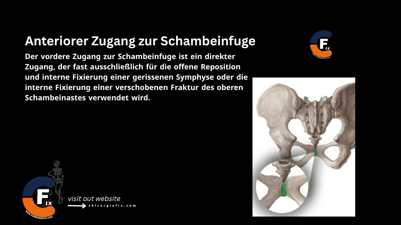
Anterior approach to the sacroiliac joint

Introduction

The anterior approach to the sacroiliac joint provides a safe and reliable way to access this structure, allowing for precise placement of anterior plates across the joint. This approach also exposes the inner wall of the pelvic bone, enabling fixation of associated pelvic fractures. Due to the joint’s anatomy, the anterior approach offers better exposure and control than the posterior approach.

Patient Positioning

  • Place the patient supine on the operating table, with a large sandbag under the buttock (the iliac crest should be elevated toward the surgeon).
  • Support the opposite pelvic wing with a padded bolster attached to the table, then tilt the table 20 degrees away from you so the mobile pelvic contents fall away.

Landmarks and Incision

Landmarks

  1. The anterior superior iliac spine
  2. The anterior third of the iliac crest

Incision

  • Make a long, curved incision over the iliac crest, starting 7 cm posterior to the anterior superior iliac spine (approximately at the level of the iliac tubercle).
  • Curve the incision forward until reaching the anterior superior iliac spine.
  • Continue the incision anteriorly and medially along the line of the inguinal ligament for another 4 to 5 cm.

Internervous Plane

  • There is no true internervous plane for the anterior approach to the sacroiliac joint. The approach simply involves stripping muscles from the pelvis; because the bone is reached via its subcutaneous surface, no muscle is denervated.

Superficial Dissection

  • Deepen the skin incision through the subcutaneous fat.
  • Expose the deep fascia overlying the gluteal muscles and the tensor fasciae latae muscle at their attachment to the outer lip of the iliac crest.
  • Next, incise the periosteum of the entire anterior third of the iliac crest, and gently strip the muscles from the outer wall of the pelvis to expose about 1 cm of the outer surface below the crest.
  • Predrill the iliac crest to allow for easy reattachment later.
  • Using an oscillating saw, cut through the wing of the ilium at this level, dividing only the outer cortex and the underlying cancellous bone.
  • Then, crack the inner cortex with an osteotome, allowing the anterior superior iliac spine—along with the divided portion of the pelvic wing—to be detached.

Deep Dissection

  • The iliacus muscle originates from the inner wall of the ilium; release it using blunt dissection.
  • As the dissection proceeds, mobilize the detached anterior superior iliac spine, which remains attached to the lateral end of the inguinal ligament.
  • This bone and muscle block must be shifted medially; to achieve this, divide some fibers of the tensor fasciae latae and sartorius muscles.
  • Strip the iliacus muscle from the inner wall of the pelvis, staying strictly in a subperiosteal plane to expose the underlying sacroiliac joint.
  • The distance is surprisingly short. As you strip the muscle, you will encounter several nutrient vessels that need to be freed from the inner pelvic wall.
  • By mobilizing the pelvic muscle from the inside of the pelvis with a large bone block, the muscles can be securely reattached later using screws during closure.

Approach Extension

  • The anterior approach to the sacroiliac joint can be extended to an ilioinguinal approach, which provides access to the entire anterior column of the acetabulum.

Dangers

Structures at risk during the anterior approach to the sacroiliac joint include:

Nerves

  1. The lateral femoral cutaneous nerve
  2. The sacral nerve roots

Vessels

  • Relatively large nutrient vessels are often avulsed from the inner wall of the ilium.

References

  • Book “Surgical Exposures in Orthopaedics” – 4th Edition
  • Campbell’s “Operative Orthopaedics,” 12th Edition

Still to read...