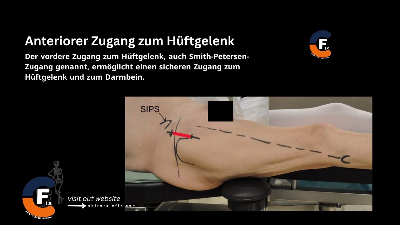
Anterior approach to the hip joint

Introduction

The anterior approach to the hip joint, also known as the Smith-Petersen approach, provides safe access to the hip joint and the ilium.

Indications

The anterior approach to the hip joint is used for:

  1. Total hip arthroplasty.
  2. Open reduction of congenital hip dislocation.
  3. Synovial biopsies.
  4. Intra-articular fusions.
  5. Resection of pelvic tumors.
  6. Irrigation and debridement of an infected native hip.
  7. The upper part of the anterior hip approach can also be used for pelvic osteotomies.

With this approach, the acetabulum is not as fully exposed as with other incisions unless the pelvic muscles are largely detached.

Patient Positioning

  • Place the patient in the supine position on the operating table.
  • If using the anterior hip approach for a pelvic osteotomy, place a small sandbag under the affected buttock to tilt the corresponding hemipelvis forward.

Landmarks and Incision

  • Landmarks:
    1. Anterior superior iliac spine (ASIS).
    2. Iliac crest.
  • Incision:
    • Make an incision from the anterior half of the iliac crest to the anterior superior iliac spine.
    • From the anterior superior iliac spine, curve the incision 8–10 cm distally toward the lateral border of the patella.

Internervous Plane

The anterior approach to the hip joint (Smith-Petersen approach) has a superficial and a deep internervous plane:

  1. The superficial internervous plane lies between:
    • Sartorius muscle: innervated by the femoral nerve.
    • Tensor fasciae latae muscle: innervated by the superior gluteal nerve.
  2. The deep internervous plane lies between:
    • Rectus femoris muscle: innervated by the femoral nerve.
    • Gluteus medius muscle: innervated by the superior gluteal nerve.

Superficial Dissection

  • Externally rotate the leg to stretch the sartorius muscle, making it more prominent and helping to identify the interval between the sartorius and tensor fasciae latae.
  • Dissect through the subcutaneous fat (take care to avoid the lateral femoral cutaneous nerve).
  • Incise the fascia on the medial side of the tensor fasciae latae muscle.
  • Release the pelvic origin of the tensor fasciae latae to develop the internervous plane.
  • The large ascending branch of the lateral femoral circumflex artery crosses the interval between the two muscles just distal to the anterior superior iliac spine. It must be ligated or coagulated.

Deep Dissection

  • Identify the plane between the rectus femoris and gluteus medius muscles.
  • Release the rectus femoris from both of its origins.
  • Retract the rectus femoris and iliopsoas medially, and the gluteus medius laterally, to expose the hip capsule.
  • Adduct and externally rotate the hip to put the capsule under tension.
  • Incise the capsule with a longitudinal or T-shaped capsulotomy.
  • Dislocate the hip by external rotation after the capsulotomy is completed.

Approach Extension

Proximal Extension

  • The anterior approach to the hip joint can be extended proximally for bone graft harvesting.
  • This is achieved by extending the proximal incision posteriorly along the iliac crest.

Distal Extension

  • The anterior approach to the hip joint can be extended distally for managing an intraoperative fracture of the distal femur.
  • Extend the skin incision distally along the anterolateral aspect of the thigh and incise the fascia lata along the line of the skin incision (stay in the interval between the vastus lateralis and rectus femoris muscles).

Dangers

Structures at risk during the anterior approach to the hip joint include:

  1. Lateral femoral cutaneous nerve:
    • The lateral femoral cutaneous nerve reaches the thigh by passing under the inguinal ligament. Its course is variable, and it may run medial or lateral to the anterior superior iliac spine.
    • It is most commonly injured when incising the fascia between the sartorius and tensor fasciae latae muscles.
    • Injury can result in a painful neuroma or diminished sensation on the lateral side of the thigh.
  2. Femoral nerve: The femoral nerve should remain protected as long as you stay lateral to the sartorius muscle.
  3. Ascending branch of the lateral femoral circumflex artery:
    • This vessel is located proximally in the internervous plane between the tensor fasciae latae and sartorius muscles.
    • Take care to ligate it to prevent excessive bleeding.

References

  • Book “Surgical Exposures in Orthopaedics” – 4th Edition
  • Campbell’s “Operative Orthopaedics”, 12th Edition

Still to read...