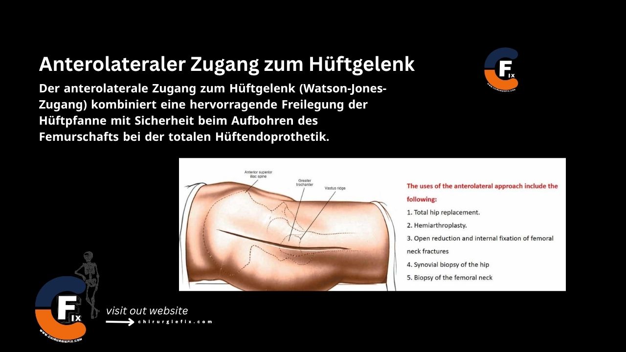
Anterolateral approach to the hip joint

Introduction

The anterolateral approach to the hip joint, also known as the Watson-Jones approach, offers excellent visualization of the acetabulum while providing safety when reaming the femoral shaft during total hip arthroplasty. This technique was popularized by Watson-Jones in 1981.

Indications for the Anterolateral Approach

  1. Total Hip Replacement (THR):
    • The minimally invasive version of this approach does not improve postoperative gait kinematics compared to the traditional transgluteal approach.
    • Patients with a high risk of dislocation may benefit from an anterolateral approach, as it avoids destruction of the posterior soft tissue.
    • Some surgeons are concerned that this approach may weaken the abductors and lead to a limp.
  2. Hemiarthroplasty.
  3. Open reduction and internal fixation of a femoral neck fracture.
  4. Synovial biopsy of the hip.
  5. Biopsy of the femoral neck.

Patient Positioning

  1. Generally, the anterolateral approach to the hip is performed with the patient in the lateral decubitus position.
  2. Alternatively, the patient can be placed supine with the buttock near the edge of the table, allowing fat to drain away from the incision.

Landmarks and Incision

Landmarks

  • Anterior superior iliac spine (ASIS).
  • Greater trochanter.
  • Shaft of the femur.
  • Vastus lateralis ridge.

Incision

  • Make the incision 2.5 cm posterior and distal to the anterior superior iliac spine (ASIS).
  • As the incision runs distally, it is centered over the tip of the greater trochanter, crosses its posterior third, and then continues along the femoral shaft.

Internervous Plane

There is no true internervous plane for the anterolateral approach to the hip joint, as the gluteus medius and tensor fasciae latae share a common nerve supply, the superior gluteal nerve.

Superficial Dissection

  • Incise the fat along the line of the skin incision and clean the fascia lata.
  • Incise the fascia: Cut in the direction of its fibers, which will be more anterior as you dissect proximally. Incise at the posterior border of the greater trochanter.
  • Develop the interval between the tensor fasciae latae and gluteus medius muscles. A small series of vessels will be found in this interval.
  • Externally rotate the hip to put the capsule under tension.
  • Identify the origin of the vastus lateralis muscle.

Deep Dissection

Release the abductor mechanism using one of two methods:

  • Trochanteric osteotomy: The distal osteotomy site is just proximal to the ridge of the vastus lateralis.
  • Partial detachment of the abductor mechanism: Place a stay suture to prevent muscle splitting and damage to the superior gluteal nerve (the nerve lies 5 cm proximal to the acetabular rim).

Expose the anterior joint capsule.

Release the reflected head of the rectus femoris from the joint capsule to expose the anterior rim of the acetabulum. This is easier when the leg is slightly flexed.

Elevate a portion of the psoas tendon from the capsule.

Perform an anterior capsulotomy.

Approach Extension

Distal Extension of the Anterolateral Approach

  • Extend the skin incision along the lateral aspect of the thigh and incise the deep fascia along the skin incision.
  • Split the vastus lateralis to gain access to the lateral part of the femur. This allows you to usefully extend the approach along the entire length of the femur.
  • A distal extension is often required when the approach is used for open reduction and internal fixation of femoral neck fractures.

Proximal Extension

  • The anterolateral approach to the hip cannot be usefully extended proximally.

Dangers

Structures at risk during the anterolateral approach to the hip include:

Femoral Nerve

  • The most common problem is compression neurapraxia caused by medial retraction.
  • Direct injury can occur from placing a retractor into the psoas muscle.

Femoral Artery and Vein

  • Can be damaged by retractors that penetrate the psoas.
  • Ensure the anterior retractor is placed directly on the bone.

Hip Abductors

  • Caused by trochanteric osteotomy and/or disruption of the abductor mechanism.
  • Caused by denervation of the tensor fasciae latae due to aggressive muscle splitting.

Femoral Shaft Fractures

  • Usually occurs during dislocation (ensure an adequate capsulotomy is performed).

References

  • Book “Surgical Exposures in Orthopaedics” – 4th Edition
  • Campbell’s “Operative Orthopaedics,” 12th Edition
  • Lepri AC, Villano M, Matassi F, Carulli C, Innocenti M, Civinini R. “Anterolateral” approach to the hip: a systematic review of the correct definition of terms. Hip Int. 2020 Dec;30(2_suppl):13-19. doi: 10.1177/1120700020966800. PMID: 33267690.

Still to read...