Introduction
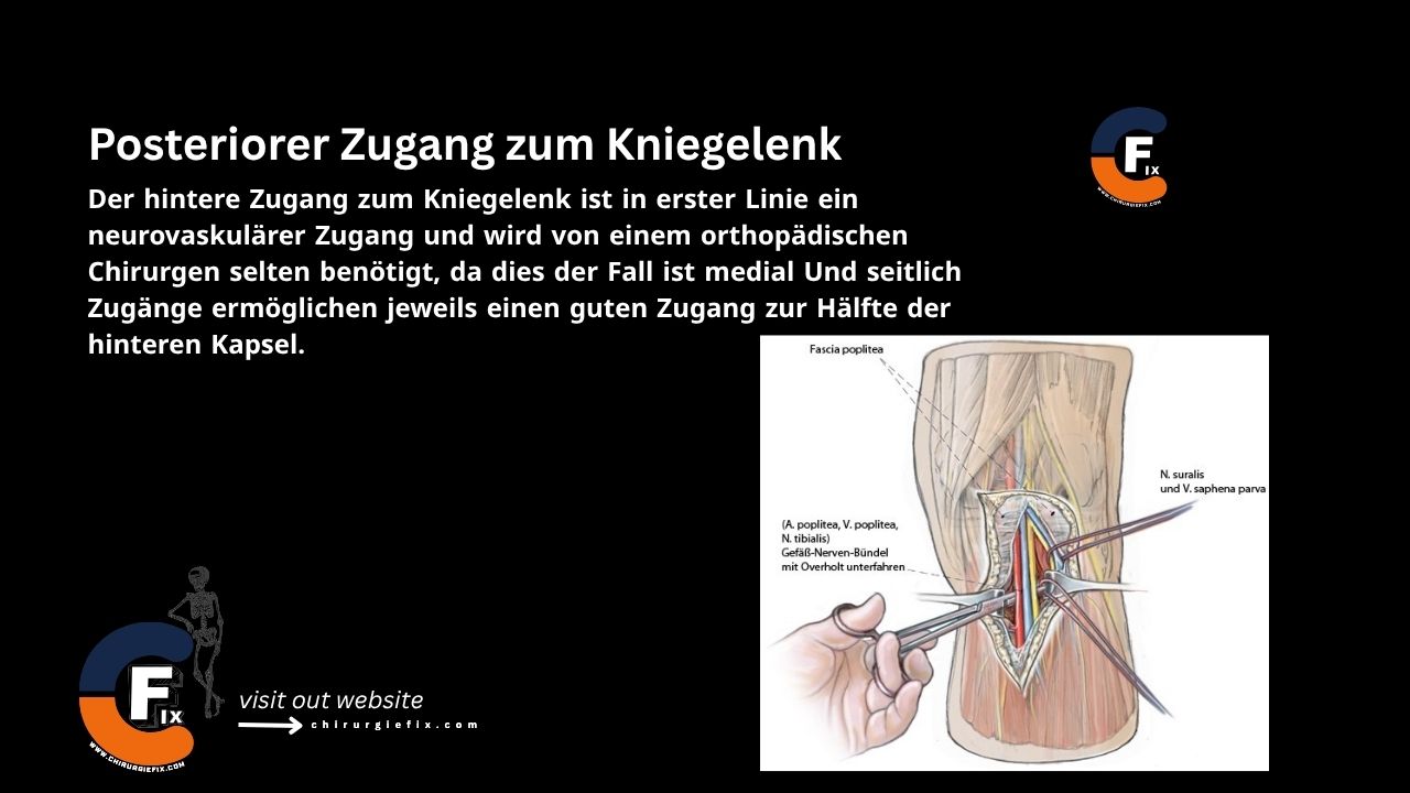
The posterior approach to the knee joint is primarily a neurovascular approach. An orthopedic surgeon rarely needs it because the medial and lateral approaches each provide excellent access to their respective halves of the posterior capsule.
Indications for the Posterior Approach
This approach is used for the following procedures:
- Repair of neurovascular structures in the popliteal fossa.
- Repair of avulsion fractures of the posterior cruciate ligament (tibial attachment).
- Recession of the gastrocnemius muscle heads for contracture.
- Lengthening of the hamstring tendons.
- Removal of Baker’s cysts and other popliteal cysts.
- Access to the posterior knee capsule.
Patient Positioning
Place the patient in the prone position on the operating table. Use a tourniquet for all procedures except vascular repairs.
Landmarks and Incision
Landmarks
- Gastrocnemius muscle.
- Semimembranosus and semitendinosus muscles.
Incision
Use a gently curved incision. Begin laterally over the biceps femoris muscle and carry the incision obliquely across the popliteal fossa. Curve it down over the medial head of the gastrocnemius and extend it into the calf.
Internervous Plane
There is no true internervous plane. This dissection exposes the contents of the popliteal fossa by incising the overlying deep fascia and retracting the three muscles that form its boundaries.
Superficial Dissection
- Reflect the skin flaps with the underlying subcutaneous fat.
- The vein is easier to identify if the leg is not fully exsanguinated before applying the tourniquet. The medial sural cutaneous nerve runs on the lateral side of the vein. The small saphenous vein can serve as a guide for the nerve, and the nerve can guide the dissection of the popliteal fossa. The nerve, continuing under the deep fascia of the calf, is a branch of the tibial nerve.
- Incise the fascia of the popliteal fossa directly medial to the small saphenous vein.
- Trace the medial sural cutaneous nerve proximally to its origin, the tibial nerve.
- Dissect to the apex of the popliteal fossa, following the tibial nerve.
- The apex of the popliteal fossa is formed medially by the semimembranosus muscle and laterally by the biceps femoris muscle.
- Near the apex, the common peroneal nerve separates from the tibial nerve.
- Dissect the common peroneal nerve from proximal to distal as it runs along the posterior border of the biceps femoris muscle.
- Now, address the popliteal artery and vein, which lie deep and medial to the tibial nerve.
- The artery has five genicular branches around the knee: two superior, two inferior, and one middle.
- If the artery needs to be mobilized, one or more of these branches may need to be ligated.
- The popliteal vein lies medial to the artery and enters the popliteal fossa from below.
- It then curves to lie directly posterior to the artery within the fossa.
- Above the knee joint, it moves to the posterolateral side of the artery.
- Be very careful when mobilizing this structure. Intimal damage can lead to thrombosis.
Deep Dissection
Retracting the muscles that form the boundaries of the popliteal fossa exposes various parts of the posterior joint capsule.
Two maneuvers can provide better access to the joint if needed:
- Posteromedial Joint Capsule: Detach the tendinous origin of the medial head of the gastrocnemius from the posterior femur. Retract the head laterally and distally, moving the nerves and vessels out of the way to reach the posteromedial corner of the joint. The exposure is now the same as that achieved by the posterior extension of the medial approach to the knee.
- Posterolateral Joint Corner: Detach the origin of the lateral head of the gastrocnemius from the lateral femoral condyle. Develop the interval between it and the biceps femoris muscle, creating the same exposure as the lateral approach to the knee.
Note that the posterior approach is not superior to the medial and lateral approaches for treating pathology in the posteromedial and posterolateral corners of the knee. It should be used mainly for exploring structures within the popliteal fossa and for reattaching the avulsed tibial insertion of the posterior cruciate ligament.
Approach Extension
- If additional medial access is needed, the medial head of the gastrocnemius can be released.
- The posterolateral extension of the posterior approach should be limited due to the risk of common peroneal nerve injury.
Dangers
Structures at risk during the posterior approach to the knee include:
- Popliteal Artery: The risk is minimized by keeping the dissection deep to the gastrocnemius muscle.
- Tibial Nerve: The risk of injury is minimized by keeping the dissection deep to the gastrocnemius muscle.
- Sural Nerve and Small Saphenous Vein.
References
- Book: “Surgical Exposures in Orthopaedics” – 4th Edition
- Book: Campbell’s “Operative Orthopaedics”, 12th Edition