Posteromedial approach to the ankle joint

Introduction

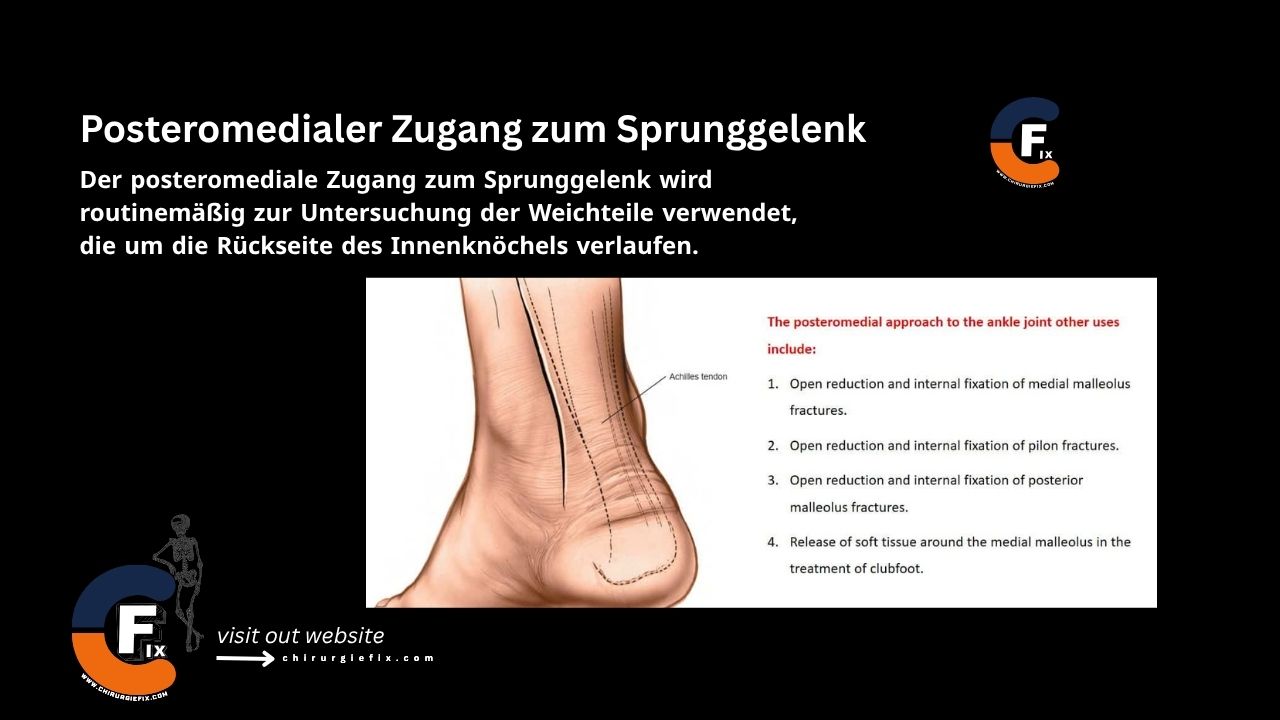
The posteromedial approach to the ankle joint is routinely used to examine the soft tissues that run around the back of the medial malleolus (the inner ankle bone).

Indications

Additional applications of the posteromedial approach include:

  1. Open reduction and internal fixation of medial malleolus fractures.
  2. Open reduction and internal fixation of pilon fractures.
  3. Open reduction and internal fixation of posterior malleolus fractures.
  4. Soft tissue release around the medial malleolus in the treatment of clubfoot.

Patient Positioning

This approach uses one of two positions:

  • Place the patient supine on the operating table. Flex the hip and knee, and rest the outer side of the affected ankle on the front of the opposite knee.
  • Place the patient in the lateral decubitus position with the affected leg closest to the table. Flex the knee of the opposite limb to move its ankle out of the way.

Landmarks and Incision

  • Landmarks:
    1. Medial malleolus.
    2. Achilles tendon.
  • Incision: Make a 10 cm curved longitudinal incision, with the concavity of the incision facing anteriorly:
    • Begin 5 cm proximal to the medial malleolus at the posterior border of the tibia.
    • Curve the incision distally along the posterior border of the medial malleolus.
    • End the incision 5 cm distal to the medial malleolus.

Internervous Plane

  • There is no internervous plane for the posteromedial approach to the ankle joint.
  • The approach is performed between:
    • Tibialis posterior tendon.
    • Flexor muscle.
      • Both are innervated by the tibial nerve.

Superficial Dissection

  • Deepen the incision along the skin incision to enter the fat that lies between the Achilles tendon and the structures running around the back of the medial malleolus. If the Achilles tendon requires lengthening, identify it in the posterior skin flap and perform the lengthening at this point.
  • Identify a fascial plane in the anterior flap that covers the remaining flexor tendons. Incise the fascia longitudinally, staying well away from the back of the medial malleolus.

Deep Dissection

There are three distinct ways to approach the back of the ankle joint:

  1. First: Identify the flexor hallucis longus, the only muscle at this level that still has muscle fibers. Develop a plane at its lateral border between it and the peroneal tendons, which lie directly lateral to it. Deepen this plane to expose the posterior part of the ankle joint by retracting the flexor hallucis longus medially.
  2. Second: Identify the flexor hallucis longus and continue the dissection anteriorly toward the back of the medial malleolus. Preserve the neurovascular bundle by carefully mobilizing it and retracting it laterally along with the flexor hallucis longus to create a plane between the bundle and the tendon of the flexor digitorum longus. This approach provides a more medial exposure of the posterior ankle joint compared to the first method.
  3. Third: If all tendons running around the back of the medial malleolus (tibialis posterior, flexor digitorum longus, and flexor hallucis longus) require lengthening, the back of the ankle can be approached directly, as the posterior tendon sheaths must be divided for the lengthening procedure. In all three methods, complete the approach by incising the joint capsule either longitudinally or transversely.

Approach Extension

  • The posteromedial approach to the ankle joint can be extended distally by curving it over the medial border of the ankle and ending it over the talonavicular joint.
  • This extension provides exposure of both the talonavicular joint and the master knot of Henry.

Dangers

Structures at risk during the posteromedial approach to the ankle joint include:

  1. Tibialis posterior muscle.
  2. Tendon of the flexor digitorum longus muscle.
  3. Tendon of the flexor hallucis longus muscle.
  4. Posterior tibial artery and vein.
  5. Tibial nerve.

References

  • Book “Surgical Exposures in Orthopaedics” – 4th edition
  • Campbell’s “Operative Orthopaedics”, 12th edition

Still to read...