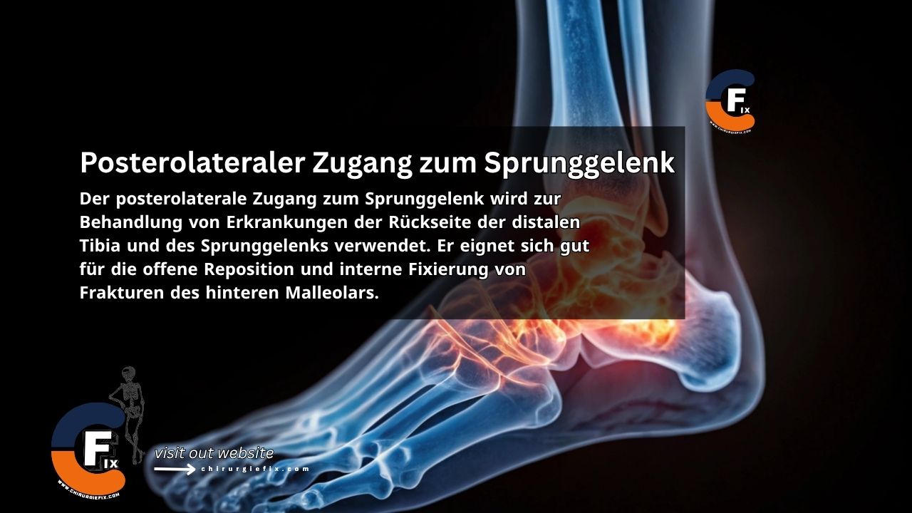
Posterolateral approach to the ankle joint

The posterolateral approach to the ankle joint is used to treat conditions affecting the posterior aspect of the distal tibia and the ankle joint. It is particularly well-suited for open reduction and internal fixation of posterior malleolar fractures.

Additional Indications

Other common uses for the posterolateral approach include:

  1. Sequestrectomy (removal of dead bone)
  2. Excision of benign tumors
  3. Arthrodesis of the posterior facet of the subtalar joint
  4. Posterior capsulotomy and syndesmotomy of the ankle
  5. Tendon lengthening procedures

However, because the patient is positioned prone, this approach is not ideal when both the fibula and the medial malleolus require simultaneous fixation. In such cases, it is preferable to use either a posteromedial or a lateral approach to the fibula, approaching the posterolateral corner of the tibia through the fibular fracture site. Neither of these alternatives offers the same degree of bone visualization as the posterolateral approach to the ankle, but both allow for the performance of other surgical procedures without needing to reposition the patient during surgery.

Patient Positioning

  • Place the patient in a prone position on the operating table.
  • Position a sandbag under the ankle to allow for dorsiflexion during the procedure.

Landmarks and Incision

  • Landmarks:
    1. Lateral malleolus
    2. Achilles tendon
  • Incision:
    • Make a 10 cm longitudinal incision halfway between the posterior border of the lateral malleolus and the lateral edge of the Achilles tendon.
    • Begin the incision at the level of the tip of the fibula and extend it proximally.

Internervous Plane

The internervous plane for the posterolateral approach to the ankle lies between:

  • Peroneus brevis muscle: innervated by the superficial peroneal nerve.
  • Flexor hallucis longus muscle: innervated by the tibial nerve.

Superficial Dissection

  • Mobilize the skin flaps. The small saphenous vein and the sural nerve run just behind the lateral malleolus.
  • Incise the deep fascia of the leg along the line of the skin incision. Identify the two peroneal tendons, which run along the leg and curve around the back of the lateral malleolus.
    • At the level of the ankle joint, the peroneus brevis tendon lies anterior to the peroneus longus tendon, and is therefore closer to the lateral malleolus.
  • Incise the peroneal retinaculum to release the tendons. Retract the muscles laterally and anteriorly to expose the flexor hallucis longus muscle.

Deep Dissection

  • Make a longitudinal incision through the lateral fibers of the flexor hallucis longus muscle where they originate from the fibula.
  • Retract the flexor hallucis longus muscle medially to expose the periosteum over the posterior aspect of the tibia.
  • If access to the distal tibia is required, develop an epiperiosteal plane between the periosteum covering the tibia and the overlying soft tissues.
  • To enter the ankle joint, follow the posterior surface of the tibia down to the posterior ankle capsule and incise it transversely.

Approach Extension

  • To extend the posterolateral approach proximally, lengthen the skin incision upward and identify the plane between the lateral head of the gastrocnemius muscle and the peroneal muscles.
  • Develop this plane down to the soleus muscle and retract it medially together with the gastrocnemius. Then, reflect the flexor hallucis longus muscle medially, releasing it from its origin on the fibula.
  • Continue the dissection medially across the interosseous membrane to reach the posterior surface of the tibia.

Dangers

Structures at risk during the posterolateral approach to the ankle include:

  • The short saphenous vein
  • The sural nerve

Both structures run close together and should be preserved as a unit, primarily to prevent the formation of a painful neuroma.

References

Still to read...