Boyd amputation

The Boyd amputation is a hindfoot-level surgical technique that preserves the calcaneus (heel bone) and fuses it with the distal tibia to create an ankle arthrodesis. This approach provides an excellent weight-bearing residual limb, often eliminating the need for a prosthetic device. However, its use is somewhat limited due to the challenges of achieving a high rate of successful union during the tibiocalcaneal fusion.

While more technically demanding than a Syme amputation, the Boyd procedure offers distinct advantages. It results in a more stable stump by preserving the function of the plantar heel pad. Furthermore, because a portion of the heel bone is retained and fused to the tibia, the weight-bearing surface is more robust compared to a Syme amputation.

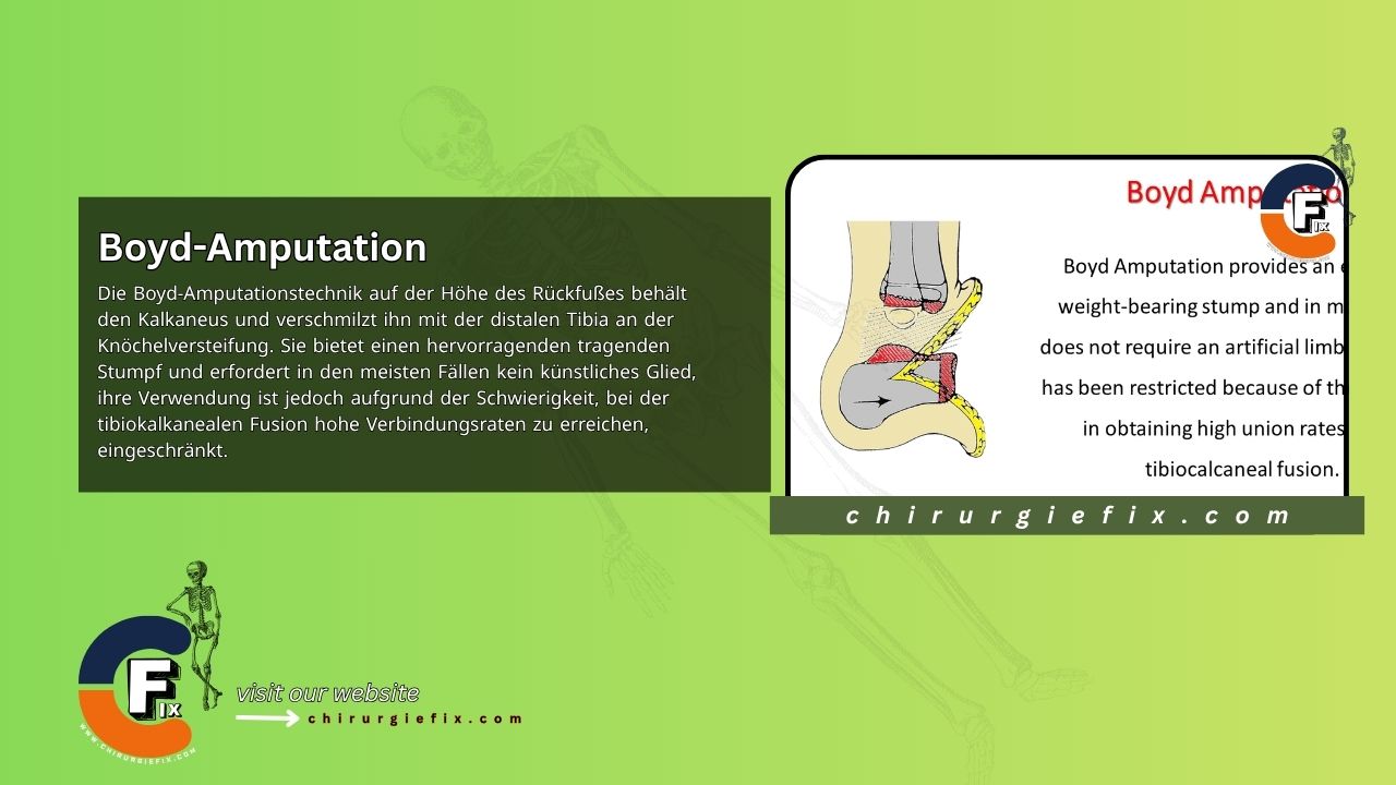
Steps of the Boyd Amputation Technique

  • Create the skin flaps: Form a long plantar flap and a shorter dorsal flap. Begin the incision at the tip of the lateral malleolus, extend across the top of the foot at the level of the talonavicular joint, and end one finger-breadth below the medial malleolus. Carry the incision down and distally along the sole of the foot, aligning it with the metatarsals. Finally, bring the incision up and proximally back to the tip of the lateral malleolus.
  • Amputate the forefoot: Lift the skin flaps and amputate the forefoot by disarticulating through the tarsometatarsal joints.
  • Remove the talus: Using precise dissection close to the bone, transect the ligaments connecting the calcaneus and tibia, then remove the talus.
  • Osteotomize the calcaneus: Perform a transverse osteotomy just below the peroneal tubercle to resect the anterior portion of the calcaneus.
  • Prepare for fusion: Remove the cartilage from the corresponding surfaces of the tibia, fibula, and calcaneus to prepare them for arthrodesis.
  • Manage tendons: Pull any visible tendons in the wound distally, then cut them proximally so they retract.
  • Address nerves: To prevent pressure-related pain, transect the medial and lateral plantar nerves.
  • Align the bones: Position the calcaneus anteriorly relative to the ankle joint and align it correctly for arthrodesis, ensuring its bottom surface remains parallel to the ground.
  • Fixate the fusion: If needed, insert a Steinmann pin or a cannulated screw from the heel upward to securely fix the calcaneus to the tibia.
  • Close the wound: Close the skin flaps with interrupted sutures and place a drain, which should be removed within 48 to 72 hours.

Post-Operative Care

  • Sutures are removed after 2 weeks; any Steinmann pins are removed after 4 weeks.
  • Weight-bearing on the stump is prohibited until the 8th week.
  • After this period, a walking cast is applied and worn until the arthrodesis is complete.

References and Further Reading

  1. Mongon ML, Sposito AL, Nunes GM, Livani B, Belangero W. Boyd Amputation with Tension Band Technique. Strategies Trauma Limb Reconstr. 2019 May-Aug;14(2):102-105. doi: 10.5005/jp-journals-10080-1433. PMID: 32742422; PMCID: PMC7376589.
  2. Grady JF, Winters CL. The Boyd Amputation for the Treatment of Osteomyelitis of the Foot. J Am Podiatr Med Assoc. 2000 May;90(5):234-9. doi: 10.7547/87507315-90-5-234. PMID: 10833871.
  3. Campbell's Operative Orthopaedics, 12th Edition.

Still to read...