Anterior approach to the ankle joint

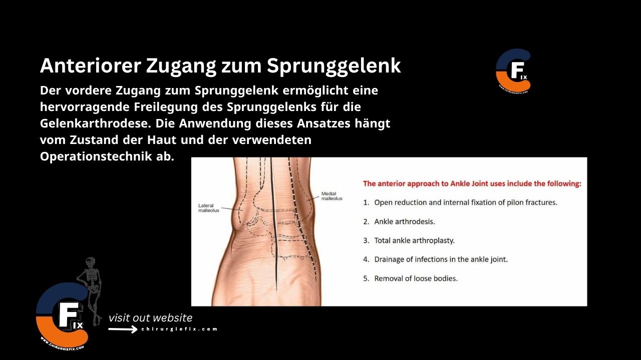
The anterior approach to the ankle joint provides excellent exposure for procedures such as arthrodesis. The choice of this approach depends on the condition of the skin and the specific surgical technique being used.

Indications for the anterior ankle approach include:

  1. Open reduction and internal fixation of pilon fractures.
  2. Ankle arthrodesis.
  3. Total ankle replacement.
  4. Drainage of ankle joint infections.
  5. Removal of loose bodies.

Patient Positioning

The patient is placed in a supine position on the operating table.

Landmarks and Incision

Landmarks

  • Medial malleolus: The subcutaneous distal end of the medial surface of the tibia.
  • Lateral malleolus: The subcutaneous distal end of the fibula.

Incision

  • Make a 15 cm longitudinal incision over the front of the ankle joint.
  • Begin the incision approximately 10 cm proximal to the joint. Extend it so that it crosses the joint roughly midway between the malleoli and ends on the dorsum of the foot. Take care to cut only the skin, as the anterior neurovascular bundle and branches of the superficial peroneal nerve cross the ankle joint very close to the skin incision line.
  • Alternatively, a 15 cm longitudinal incision can be made, centered over the anterior part of the medial malleolus.

Internervous Plane

  • The anterior approach to the ankle does not have a true internervous plane.
  • The muscles of the extensor hallucis longus and extensor digitorum longus define a clear intermuscular plane. Both muscles are supplied by the deep peroneal nerve; however, the plane can be used because both receive their nerve supply well proximal to the dissection level.
  • This plane must be used with great caution as it contains the neurovascular bundle distal to the ankle.

Superficial Dissection

  • Incise the deep fascia of the leg along the skin incision, cutting through the extensor retinaculum.
  • Identify the plane between the extensor hallucis longus and extensor digitorum longus muscles a few centimeters above the ankle joint. Locate the neurovascular bundle (anterior tibial artery and deep peroneal nerve) just medial to the tendon of the extensor hallucis longus.
  • Trace the bundle distally until it crosses the front of the ankle joint behind the extensor hallucis longus tendon.
  • Retract the extensor hallucis longus tendon medially along with the neurovascular bundle.
  • Retract the extensor digitorum longus tendon laterally. The tendons become mobile after the retinaculum is divided, but the neurovascular bundle is adherent to the underlying tissue and must be mobilized.
  • Alternatively, for pilon fractures, the deep fascia can be incised on the medial side of the tibialis anterior tendon to expose the underlying surface of the tibia along with the anteromedial ankle capsule.

Deep Dissection

  • For an ankle arthrodesis using the anterior approach, incise the remaining soft tissue longitudinally to expose the anterior surface of the distal tibia.
  • Continue the incision down to the ankle joint and then divide the anterior capsule. Expose the full width of the ankle joint by detaching the anterior capsule from the tibia or talus using sharp dissection. Some periosteal elevation from the distal tibia may be necessary.
  • While the periosteal layer is usually thick and easy to define, the plane may be obliterated in cases of infection. In such cases, the periosteum must be detached piece by piece using sharp dissection.
  • When using this approach for fracture surgery, take particular care to preserve as many soft tissue attachments to the bone as possible.
  • Careful preoperative planning allows for smaller, more precise incisions, which reduces soft tissue damage.

Approach Extension

  • The anterior approach to the ankle can be extended proximally to expose structures in the anterior compartment.
  • To expose the proximal tibia, use the plane between the tibia and the tibialis anterior muscle.
  • Distal extension onto the dorsum of the foot is possible but rarely required.

Dangers

Structures at risk during the anterior ankle approach include:

  1. Superficial cutaneous branches of the peroneal nerve: These are most at risk during the skin incision.
  2. Neurovascular bundle (deep peroneal nerve and anterior tibial artery): Above the joint, it runs between the extensor digitorum longus and extensor hallucis longus, crossing behind the extensor hallucis longus at the level of the joint.

References and Further Reading

  1. Surgical Exposures in Orthopaedics – 4th Edition
  2. Campbell's Operative Orthopaedics, 12th Edition
  3. Dekker RG 2nd, Kadakia AR. Anterior Approach to Ankle Arthrodesis. JBJS Essent Surg Tech. 2017 Apr 12;7(2):e1. DOI: 10.2106/JBJS.ST.15.0. PMID: 30233945; PMCID: PMC6132605.
  4. World J Orthop. 2014 Jan 18;5(1):1-5, Foot Ankle Int. 2011 Oct;32(10):940-7, and Foot Ankle Int. 2009 Jul;30(7):631-9.

Still to read...