Arthroscopic knee synovectomy is a safe and effective procedure that offers significant potential advantages over open surgical techniques. These benefits include improved visualization of the knee joint, a more complete synovectomy, less postoperative pain, reduced knee stiffness and arthrofibrosis, decreased postoperative hemarthrosis, a shorter hospital stay, lower morbidity at the surgical site, and the ability to perform the operation through small arthroscopic portals (which avoids splitting the quadriceps muscle). This technique also allows for better preservation of the menisci, and any necessary revision surgery is less complicated.
The main arthroscopic disadvantage of knee synovectomy is that the procedure can be technically more demanding and is contraindicated for some diffuse and malignant conditions.
The knee joint has the largest and most extensive synovial membrane of any joint in the body. As a result, pathological conditions affecting the knee's synovium can be both symptomatic and debilitating. Both benign and malignant processes can involve the synovial membrane.
For intra-articular malignancies (e.g., synovial sarcoma), treatment typically involves radical excision combined with chemotherapy and/or radiation therapy.
Lack of surgeon experience or skill: Performing a thorough synovectomy requires excellent technical skills and knowledge of arthroscopy. An inadequate or incomplete procedure can lead to local recurrence and poor outcomes. Therefore, arthroscopic synovectomy is not recommended if the surgeon is not confident in performing a complete and adequate procedure. In such cases, a traditional open approach should be chosen, or the patient should be referred to an experienced arthroscopist.
Severe arthritic changes, deformities, or contractures: These conditions can include advanced osteoarthritis, rheumatoid arthritis, seronegative arthritis, and hemophilia. In such cases, total knee arthroplasty is the definitive treatment of choice.
The patient is placed supine on the operating table. It is preferred that the operative leg is placed in a thigh holder positioned four finger-breadths above the superior pole of the patella. This allows the leg to hang freely off the end of the table when the foot of the bed is lowered, enabling deep flexion and varus/valgus stress without compromising portal placement. The non-operative leg is placed in a leg holder.
Alternatively, arthroscopic synovectomy can be performed with the patient supine, the knee flexed over the side of the table against a lateral post, and the non-operative leg lying flat on the operating table. The operative extremity is then prepped and draped in the usual sterile fashion.
Correct portal placement is critical for any arthroscopic procedure. Improper portal placement can lead to iatrogenic injury to the knee and an inadequate synovectomy due to procedural difficulty. It is often helpful to draw the anatomy before creating the portals.
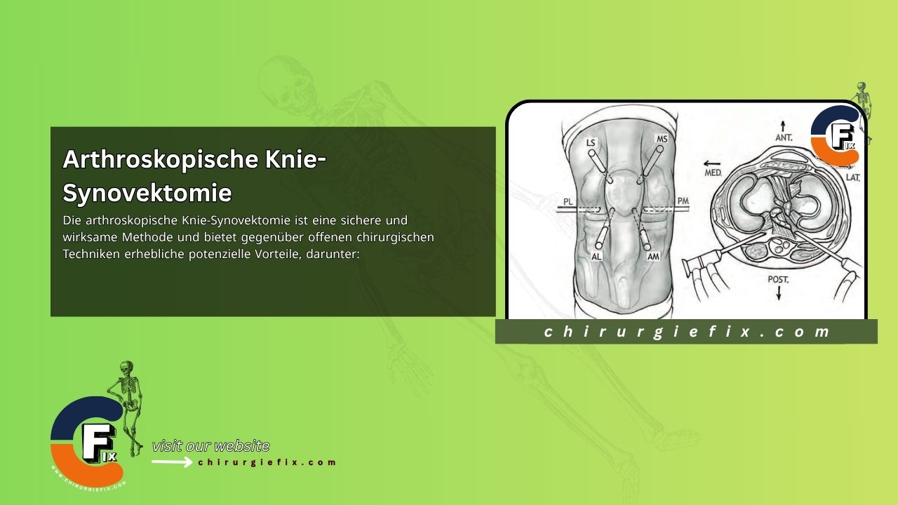
A complete arthroscopic synovectomy of the knee can be performed using a combination of six portals: anterolateral, anteromedial, lateral suprapatellar, medial suprapatellar, posterolateral, and posteromedial.
After establishing the anterolateral and anteromedial portals, a standard diagnostic arthroscopy is performed. This involves visualizing and examining the suprapatellar pouch, the medial and lateral gutters, the trochlear groove, the undersurface of the patella, the medial and lateral compartments (including the menisci), the intercondylar notch, and the cruciate ligaments. Any concurrent intra-articular pathology observed during this exam, such as meniscal or cartilage injuries, can be addressed throughout the procedure. If a pathological specimen is required, an arthroscopic grasper can be used through the anteromedial portal to obtain a synovial tissue sample from an area of significant pathology. Alternatively, an arthroscopic trap can be placed in the shaver's suction line to collect a substantial amount of resected synovium.
With the camera viewing the suprapatellar pouch, the lateral and medial suprapatellar portals are established under direct vision. These are placed approximately 1 cm above and 1 cm lateral (for the lateral suprapatellar portal) or medial (for the medial suprapatellar portal) to the corner of the patella. A 5.5-mm or 4.5-mm full-radius synovectomy blade can be used through these portals to resect the synovium in the suprapatellar pouch and the superior lateral and medial gutters, as well as to access any part of a hypertrophic anterior fat pad. Resection is sufficient when the shiny capsular layer directly underneath is visible. The lower portions of the lateral and medial gutters can be accessed for resection by alternating the camera through the lateral and medial suprapatellar portals while using the shaver through the anterolateral and anteromedial portals. Synovectomy in the anterior compartment and the intercondylar notch can then be achieved by triangulating the arthroscope and shaver through the anteromedial and anterolateral portals. These two portals can also be used to visualize and resect the inferior lateral and medial gutters.
For safe posterior knee arthroscopy, there is a 15- to 28-mm safety zone between the posterior cruciate ligament and the popliteal neurovascular bundle. Keeping the knee flexed to 70–90 degrees improves access to the posterior compartment by widening the intercondylar notch and allowing the neurovascular bundle to fall further posteriorly. A 70-degree arthroscope is then used to visualize the posterior compartment. This also allows the posterior neurovascular bundle to fall away posteriorly.
A modified Gillquist maneuver is performed with the arthroscope through the anterolateral portal, advancing it under the posterior cruciate ligament to establish a posteromedial portal under direct visualization. Palpation of the posteromedial knee is performed under visualization to better locate the portal site. At this point, a spinal needle is introduced into the posteromedial corner of the knee joint, aiming anteriorly. Dimming the overhead and room lights and using transillumination from the arthroscope can also be helpful when introducing the spinal needle.
The posteromedial portal is then created, and a blunt cannula is inserted through it. The cannula is best inserted over a switching stick for easier and more accurate placement. Caution is necessary when introducing any instrument into the joint at this location, with emphasis on aiming slightly anterior to avoid iatrogenic injury to the posterior neurovascular bundle. The posteromedial portal is typically placed 16 to 35 mm from the saphenous vein.
Next, an arthroscopic synovectomy of the posteromedial knee is performed systematically from the periphery to the center through this portal using a 4.5-mm shaver. During this part of the procedure, careful attention must be paid to suction to avoid pulling the posterior capsule into the shaver, which could risk accidental iatrogenic injury to vital posterior structures.
Finally, the lateral portion of the posterior compartment is accessed in a similar manner by shifting the arthroscope to the anteromedial portal. Flexing the knee to 90 degrees allows the common peroneal nerve to fall posteriorly behind the biceps femoris tendon. A spinal needle is introduced anterior to the biceps femoris tendon, 1 cm above the joint line and 1 cm behind the femoral condyle. The posterolateral portal is created and then cannulated like the posteromedial portal. The posterolateral portal is typically placed 40 to 52 mm from the peroneal nerve. The synovectomy of the posterolateral knee is then performed systematically, similar to the posteromedial side.
Depending on the extent of the arthroscopic synovectomy, a drain may be placed and monitored for at least one day before removal to reduce postoperative hemarthrosis. This may not be necessary if bleeding is not significant at the conclusion of the case. A compression bandage is applied to the knee. Patients may be allowed partial weight-bearing with crutches and are typically discharged on the same day or the first postoperative day.
Cryotherapy with ice packs can help provide postoperative pain and swelling relief. Oral pain medication should be prescribed, and physical therapy should begin early to ensure a quick return to normal gait, range of motion, and strength.
Patients should be given written home instructions so they can begin a postoperative home therapy program immediately, focusing on active range of motion and quadriceps strengthening. Although not critical, the use of a continuous passive motion (CPM) machine is helpful to facilitate early motion. The majority of patients achieve near-normal activity levels by the fourth postoperative week.
The complications of arthroscopic synovectomy are similar to those of any arthroscopic procedure, including anesthesia-related issues, infection, bleeding, deep vein thrombosis, arthrofibrosis, and iatrogenic damage to articular cartilage or neurovascular structures.
During the procedure, care must be taken when maneuvering the arthroscope or instruments to avoid excessive pressure on the articular cartilage. Instruments should be kept away from at-risk posterior structures when using the posterolateral and posteromedial portals, namely the neurovascular bundle, the saphenous vein and saphenous nerve, and the common peroneal nerve.
Other complications more specific to arthroscopic synovectomy include hemarthrosis (leading to pain and stiffness), postoperative arthrofibrosis (which may require manipulation or arthroscopic lysis of adhesions), reflex sympathetic dystrophy, or incomplete resection leading to the recurrence or persistence of synovial pathology, thereby requiring additional surgical intervention.