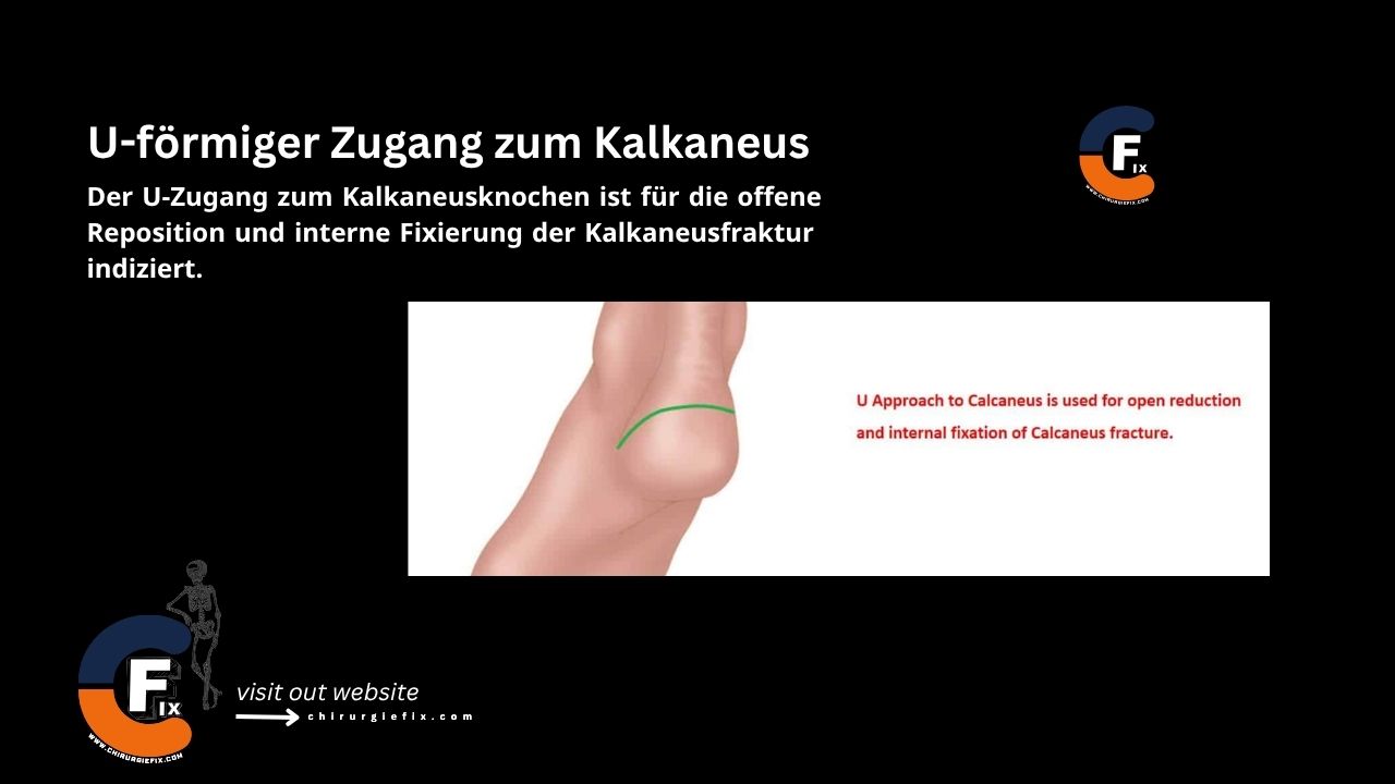
The U-shaped approach to the calcaneus is indicated for open reduction and internal fixation of calcaneus fractures.
Patient Positioning
Place the patient in the prone position on the operating table.
Support the operative leg on a sandbag.
Landmarks and Incision
Make a lateral incision as described in the lateral approach to the calcaneus.
Make a medial incision as described in the medial approach to the calcaneus.
Connect the medial and lateral incisions with a transverse posterior incision below the Achilles tendon insertion.
This creates a U-shaped incision around the posterior four-fifths of the bone.
Internervous Plane
There is no internervous plane for the U-shaped approach to the calcaneus.
Deep Dissection
Divide the superficial and deep fascia.
Incise the periosteum transversely to the line of the incision.
Perform subperiosteal dissection to create a U-shaped flap consisting of skin, the fatty heel pad, plantar aponeurosis, and muscles.
Elevate the plantar aponeurosis and the muscles of the calcaneus to expose the bone.
Dangers
Structures at risk during the U-shaped approach to the calcaneus include:
Sural nerve (lateral): Located 18.8 mm from the lateral border of the Achilles tendon at its insertion and crosses the lateral border of the Achilles tendon 9.8 cm proximal to the insertion.
Medial calcaneal nerve (medial).
Nerve to the abductor digiti minimi muscle (medial).
References
Campbell's Operative Orthopaedics, 12th edition.
Follow Us & Stay Updated!
Don't miss new scholarships, universities, orthopedic insights, physiotherapy resources, and medical education updates.