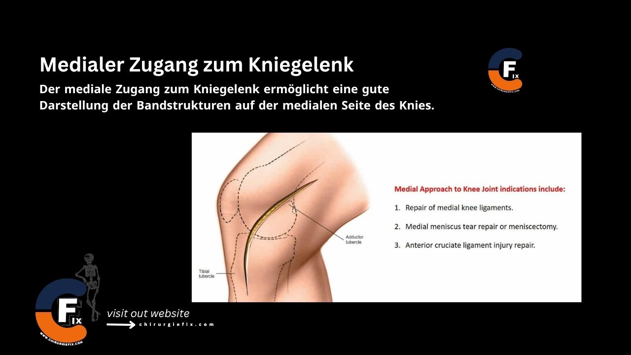
The medial approach to the knee joint provides excellent exposure of the ligamentous structures on the inner side of the knee.
The infrapatellar branch of the saphenous nerve crosses the operative field transversely and is sacrificed. However, the saphenous nerve itself, which emerges between the gracilis and sartorius muscles, must be preserved, along with the long saphenous vein in the posteromedial part of the dissection.
(The infrapatellar branch of the saphenous nerve should be divided and its end buried in fat to reduce the likelihood of forming a painful neuroma.)
To expose the deep structures of the knee, the layers covering them must be incised either in front of or behind the superficial medial collateral ligament (the medial collateral ligament).
These separate incisions allow access to the anterior and posterior parts of the medial side of the joint, respectively.
Use the anterior approach to expose the superficial medial ligament, the anterior part of the medial meniscus, and the cruciate ligament.
The posterior approach exposes the posterior third of the meniscus and the posteromedial corner of the knee.
Structures at risk during the medial approach to the knee joint include: