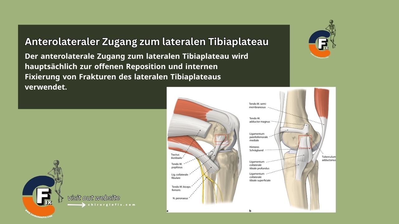
Anterolateral approach to the lateral tibial plateau

The anterolateral approach to the lateral tibial plateau is primarily used for open reduction and internal fixation of lateral tibial plateau fractures.

Other applications of the anterolateral approach to the lateral tibial plateau include:

  1. Bone grafting for delayed union and nonunion of fractures.
  2. Treatment of osteomyelitis.
  3. Tumor excision and biopsy.
  4. Proximal tibial osteotomy.
  5. Bone graft harvesting.

The anterolateral approach is preferred over a direct anterior approach to the tibia because the skin in the anterolateral approach does not lie directly over the bone.

Patient Positioning

  • Place the patient supine on a radiolucent table.
  • Position a firm wedge under the knee to flex the joint to approximately 60°.
  • Place a small bag under the ipsilateral buttock to correct the natural external rotation of the lower extremity. This ensures the patella faces directly forward.

Landmarks and Incision

  • Landmarks:
    • The shaft of the proximal tibia along its anterior border.
    • The lateral joint line.
    • Gerdy's tubercle.
  • Incision:
    • Make an S-shaped incision. Begin approximately 3 to 5 cm proximal to the joint line, staying just lateral to the border of the patellar tendon.
    • Curve the incision anteriorly over Gerdy's tubercle, then extend it distally, staying about 1 cm lateral to the anterior border of the tibia.
    • The exact length of the incision depends on the pathology being treated and the implant being used.

Internervous Plane

  • There is no true internervous plane in the anterolateral approach to the lateral plateau.
  • The dissection is essentially epiperiosteal and does not disturb the nerve supply to the extensor compartment.

Superficial Dissection

  • Deepen the incision proximally through the subcutaneous tissue to expose the lateral aspect of the knee joint capsule.
  • The knee joint capsule is incised longitudinally down to the superior border of the lateral meniscus.
  • Below the joint line, deepen the incision through the subcutaneous tissue and incise the fascia over the tibialis anterior muscle.

Deep Dissection

  • Proximal access to the knee joint is achieved by incising the synovial membrane.
  • Carefully detach the lateral meniscus from its soft tissue attachments inferiorly, developing a plane between the underside of the lateral meniscus and the underlying tibial plateau.
  • Place traction sutures on the edge of the meniscus to facilitate reattachment during closure.
  • Detach a sufficient portion of the meniscus to allow adequate visualization of the superior surface of the lateral tibial plateau.
  • Using an elevator, partially detach the origin of the tibialis anterior muscle from the proximal tibia.
  • Attempt to work in a plane between the periosteum and the muscle.

Approach Extension

Proximal Extension:

  • To extend the anterolateral approach to the lateral plateau proximally, continue the skin incision along the lateral side of the patella, then curve it posteriorly over the lateral aspect of the distal femur.
  • Deepen the incision through the lateral joint capsule to gain proximal access to the knee joint and the distal femur.

Distal Extension:

  • To extend the approach distally, continue the incision longitudinally, staying 1 cm lateral to the anterior border of the tibia.
  • Extend it proximally as far as the ankle.
  • Through deep dissection, either by splitting the tibialis anterior muscle or by detaching it from the lateral aspect of the tibia, access to the tibial shaft down to its proximal quarter is provided.

Dangers

Structures at risk during the anterolateral approach to the lateral plateau include:

  1. The superficial branch of the peroneal nerve: It lies well posterior to the dissection area and should not be injured.
  2. The lateral meniscus: Take care not to detach it completely; preserve its anterior and posterior attachments.

References

  • Surgical Exposures in Orthopaedics - 4th Edition
  • Campbell's Operative Orthopaedics, 12th Edition

Still to read...