Medial approach to the hip joint

Medial Approach to the Hip Joint

The medial approach to the hip, attributed to Ludloff, was originally developed for surgeries on hips that are flexed, abducted, and externally rotated—deformities caused by certain types of congenital hip dislocations.

Indications for the Medial Approach

This surgical approach is used for several procedures, including:

  • Open reduction of developmental dysplasia of the hip (DDH)
  • Release of the psoas tendon (this approach provides excellent exposure of the tendon)
  • Biopsy and treatment of tumors in the lower femoral neck and the medial side of the proximal femoral shaft
  • Obturator neurectomy (the upper part of this approach can be used for this purpose)

Patient Positioning

  • Place the patient supine on the operating table with the affected hip flexed, abducted, and externally rotated.
  • The sole of the foot on the affected side should rest against the medial side of the opposite knee.

Landmarks and Incision

Landmarks

  • Adductor longus muscle
  • Pubic tubercle

Incision

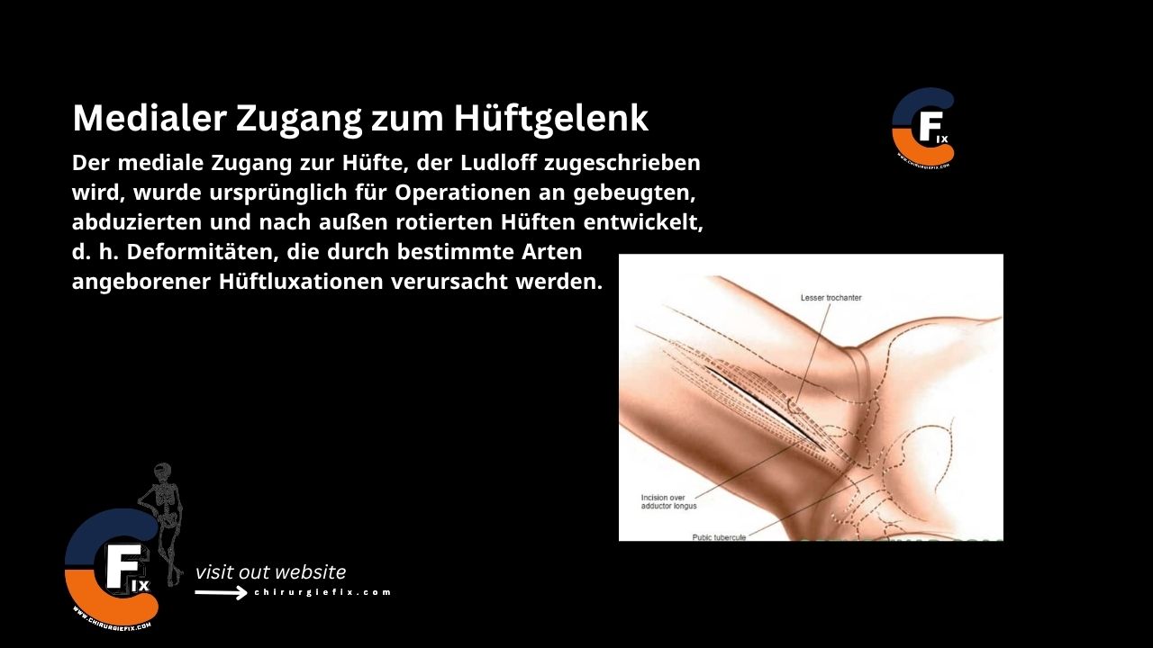
  • Make a longitudinal incision on the medial side of the thigh, starting 3 cm below the pubic tubercle.
  • The incision runs over the adductor longus muscle. Its length is determined by the amount of femur that needs to be exposed.

Internervous Plane

Superficial

There is no superficial internervous plane for the medial approach to the hip. Both the adductor longus and gracilis muscles are innervated by the anterior branch of the obturator nerve.

Deep

The deep internervous plane for the medial approach to the hip joint lies between the adductor brevis and adductor magnus muscles.

  1. The adductor brevis is supplied by the anterior branch of the obturator nerve.
  2. The adductor magnus has a dual nerve supply:
    • The adductor part is supplied by the posterior branch of the obturator nerve.
    • The hamstring part is supplied by the tibial division of the sciatic nerve.

Superficial Dissection

  • Develop a plane between the gracilis muscle and the adductor longus muscle.

Deep Dissection

  • Develop a plane between the adductor brevis and adductor magnus until you feel the lesser trochanter at the base of the wound.
  • Try to protect the posterior branch of the obturator nerve (which innervates the adductor part of the muscle) to preserve the hip extensor function of the adductor magnus.
  • Place a narrow retractor (e.g., a bone spike) above and below the lesser trochanter to isolate the psoas tendon.

Approach Extension

  • The medial approach to the hip joint cannot be extended.

Dangers

Structures at risk during the medial approach to the hip joint include:

  1. Medial femoral circumflex artery:
    • This artery courses around the medial side of the distal part of the psoas tendon.
    • In children, there is a risk during psoas tendon release. The tendon must be isolated and divided under direct vision.
  2. Anterior branch of the obturator nerve:
    • It supplies the adductor longus, adductor brevis, and gracilis muscles in the thigh.
  3. Posterior branch of the obturator nerve:
    • It lies within the substance of the obturator externus muscle.
    • It supplies the adductor part of the adductor magnus muscle.
  4. Deep external pudendal artery:
    • It is at risk proximally.
    • It lies anterior to the pectineus muscle, near the origin of the adductor longus muscle.

Related Anatomy

The adductor compartment of the thigh consists of three muscle layers. The two branches of the obturator nerve run between each pair of layers.

  1. The superficial layer is composed of the adductor longus and gracilis muscles.
  2. The middle layer is the adductor brevis muscle.
  3. The deep layer is the adductor magnus muscle.

The adductor longus is the only muscle of the adductor group that is easily palpable at its tendinous origin. Its structure is examined in detail during the superficial surgical dissection.

  • The gracilis is extremely long and thin, with long, parallel fibers. Its aponeurotic origin, a thin sheet of tendon fibers arising from the pubic bone, lies in an anteroposterior plane.
  • The adductor longus arises from a strong tendon, which explains its involvement in the relatively high incidence of avulsion fractures.
  • The obturator nerve arises from the anterior divisions of the L2–L4 nerve roots. The nerve divides into an anterior and a posterior branch in the obturator canal. The anterior branch runs over the upper border of the obturator externus muscle and descends on the medial side of the thigh behind the adductor longus, onto the anterior surface of the adductor brevis. It provides sensory fibers to the hip joint.

References

  • Book: "Surgical Exposures in Orthopaedics" – 4th Edition
  • Campbell's "Operative Orthopaedics", 12th Edition

Still to read...


























