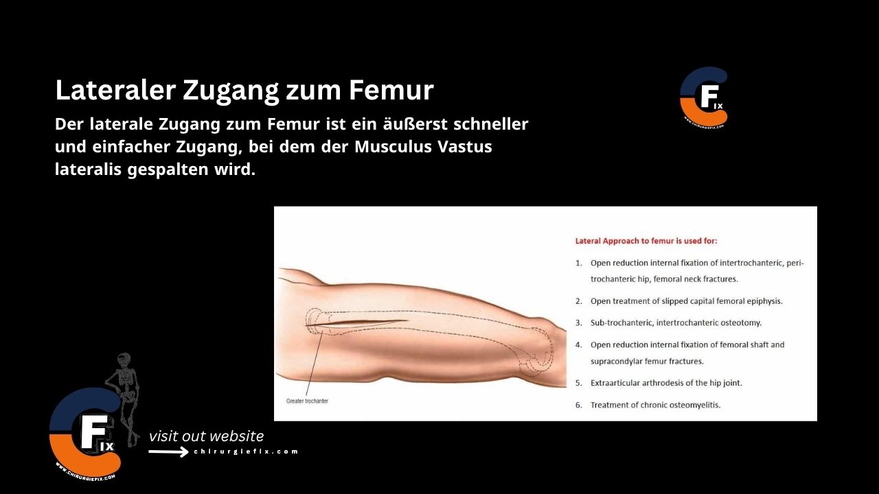
Lateral approach to the femur

The lateral approach to the femur is a fast and straightforward surgical technique that involves splitting the vastus lateralis muscle.

This approach is indicated for:

  1. Open reduction and internal fixation of intertrochanteric, pertrochanteric, and femoral neck fractures.
  2. Open treatment of a slipped capital femoral epiphysis.
  3. Subtrochanteric and intertrochanteric osteotomy.
  4. Open reduction and internal fixation of femoral shaft and supracondylar femur fractures.
  5. Extra-articular arthrodesis of the hip joint.
  6. Treatment of chronic osteomyelitis.
  7. Biopsy and treatment of bone tumors.

Blood loss from vessel disruption during this procedure can complicate the surgery, though it is rarely life-threatening.

Patient Positioning

Supine Position:

  • Place a sandbag under the buttock to internally rotate the operative leg.

Supine on an Orthopedic Table:

  • The supine position is used for trochanteric fractures.
  • Use an orthopedic table for all procedures requiring an image intensifier.

Lateral Decubitus Position:

  • This position, with the affected limb facing upward, is best for surgeries on the femoral shaft.

Landmarks and Incision

Landmarks:

  • Greater trochanter
  • Shaft of the femur

Incision:

  • Make a longitudinal incision starting at the midpoint of the greater trochanter, extending distally along the lateral aspect of the thigh over the lateral side of the femur. The length and exact location of the incision depend on the surgical requirements.

Internervous Plane

  • There is no true internervous plane for the lateral approach to the femur, as the dissection involves splitting the vastus lateralis muscle, which is innervated by the femoral nerve.
  • However, the vastus lateralis receives its nerve supply high in the thigh, so splitting the muscle distally does not lead to denervation.

Superficial Dissection

Incise the fascia lata of the thigh along the line of the skin incision. At the proximal end of the wound, you may need to split the distal part of the tensor fasciae latae muscle along its fibers to expose the vastus lateralis.

Deep Dissection

  • Carefully incise the fascial covering of the vastus lateralis muscle.
  • Insert a Hohmann or Bennett retractor through the muscle, guiding its tip over the anterior surface of the femoral shaft.
  • Then, insert a second retractor through the same split down to the femoral shaft.
  • Manipulate the second retractor so it passes beneath the femur, and pull the two retractors apart to split the vastus lateralis along its fiber line.
  • Continue splitting the muscle using blunt dissection. As you proceed, several perforating vessels crossing the field will be exposed. Coagulate them if possible before they are torn by the blunt dissection.
  • Splitting the vastus lateralis reveals the underlying lateral surface of the femur.

Extension of the Approach

  • The lateral approach is most useful for exposing the proximal third of the femur for internal fixation of hip fractures.
  • It can be extended distally to the knee joint, allowing complete exposure of the lateral femoral shaft for reduction and fixation of all types of femoral fractures.

Dangers

Structures at risk during this approach include:

  1. When dissecting the vastus lateralis, there is a risk of perforating branches of the profunda femoris artery. These should be ligated to prevent hematoma formation.

References

  • “Surgical Exposures in Orthopaedics” – 4th Edition.

Still to read...