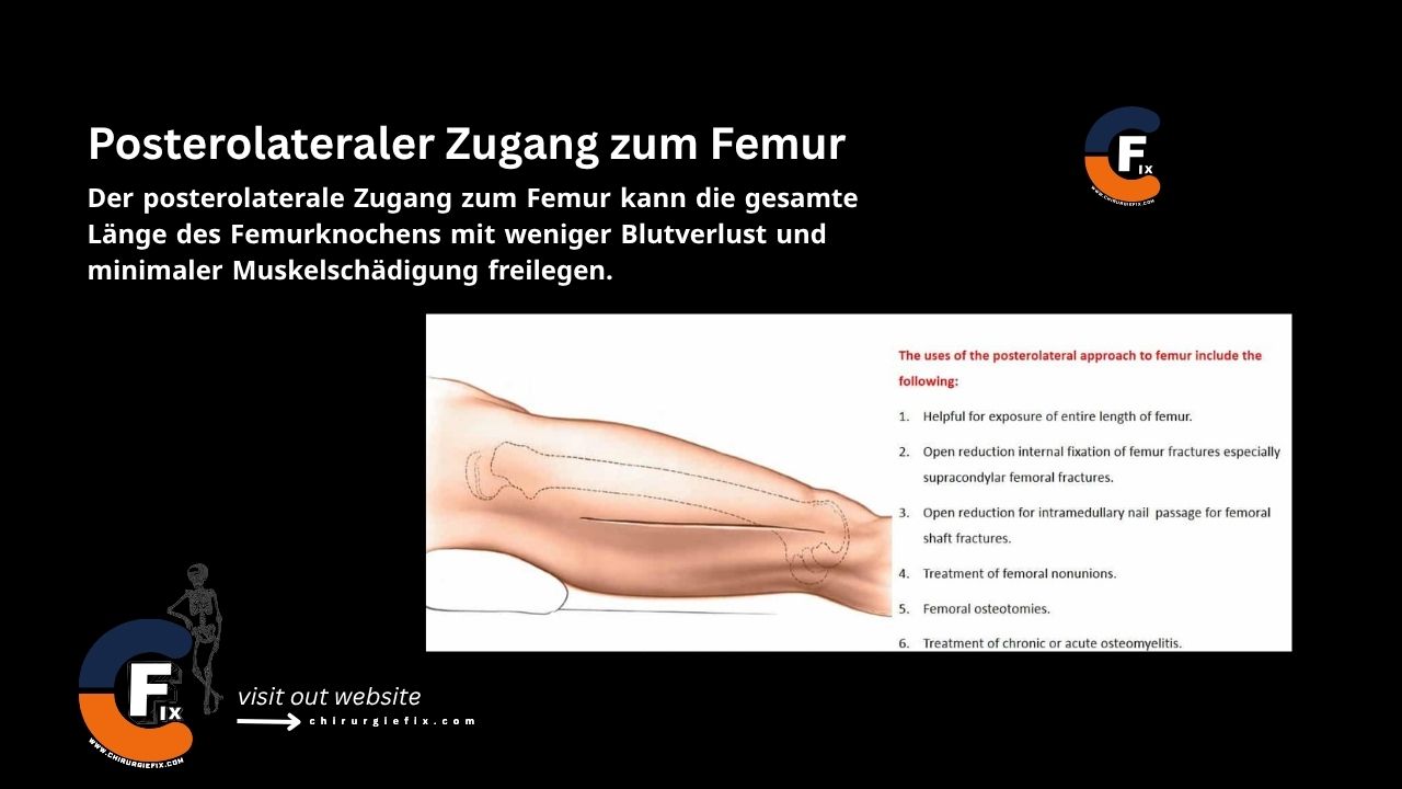
Lateral approach to the calcaneus

Introduction

The lateral approach to the calcaneus is primarily used for open reduction and internal fixation of calcaneal fractures.

Indications

Indications for the lateral approach to the calcaneus include:

  1. Open reduction and internal fixation of displaced calcaneal fractures.
  2. Treatment of other lesions of the posterior facet of the subtalar joint and the lateral wall of the calcaneus.

Calcaneal fractures are always accompanied by significant soft tissue swelling. To reduce the risk of skin necrosis, it is important to allow this soft tissue swelling to subside before surgery. A thorough assessment of the patient’s vascular status is essential prior to the operation.

Diabetes, especially when combined with neuropathy, and smoking are relative contraindications for this surgical approach.

For more details, watch this video: Calcaneus Fractures – Operative Techniques (OTA Lecture)

Patient Positioning

  • Place the patient in a lateral decubitus position on the operating table.
  • Ensure good padding over all bony prominences. Position the operative leg posteriorly with the lower leg forward.

Landmarks and Incision

  • Landmarks:
    1. The posterior border of the distal fibula.
    2. The lateral border of the Achilles tendon.
    3. The styloid process at the base of the fifth metatarsal.
  • Incision:
    • The skin incision has two limbs.
    • Begin the distal part of the incision at the base of the fifth metatarsal and extend it posteriorly, following the junction between the smooth skin of the dorsum of the foot and the wrinkled skin of the sole.
    • Make a second incision, starting approximately 6 to 8 cm above the heel skin, midway between the back of the fibula and the outer side of the Achilles tendon.
    • Extend this second incision distally until it meets the first incision, which lies over the lateral side of the calcaneus.

Internervous Plane

  • There is no internervous plane for the lateral approach to the calcaneus.
  • Dissection is performed through a direct approach to the subcutaneous bone.

Superficial Dissection

  1. Deepen the skin incision through the subcutaneous tissue, being careful not to raise any flaps.
  2. Distally, dissect straight down sharply to the lateral surface of the calcaneus.

Deep Dissection

  • Incise the periosteum of the lateral wall of the calcaneus and develop a full-thickness flap consisting of periosteum and all overlying tissues.
  • Stay on the bone and retract the soft tissue flap further proximally. The peroneal tendons are carried anteriorly with the flap.
  • Divide the calcaneofibular ligament to expose the subtalar joint.
  • Continue the dissection proximally to expose the body of the calcaneus and the subtalar joint.
  • The calcaneocuboid joint is exposed distally by incising its capsule. Take care to avoid cutting into the muscle belly of the abductor digiti minimi muscle as much as possible.

Dangers

Structures at risk during the lateral approach to the calcaneus include:

  • The sural nerve.
  • The skin:
    • The risk of skin necrosis can be minimized if the flap is raised as a full-thickness flap, as the skin receives its blood supply from the underlying tissue.

Dissecting skin flaps in this area, which is always severely traumatized, is associated with a significant number of wound complications. A thorough preoperative assessment of the patient’s vascular status is crucial. Most surgeries in this area must be postponed for an extended period to allow soft tissue swelling to subside before the operation begins.

References

  • Book “Surgical Exposures in Orthopaedics” – 4th Edition.
  • Campbell’s Book “Operative Orthopaedics”, 12th Edition.

Still to read...