Ollier approach to the sinus tarsi

Introduction

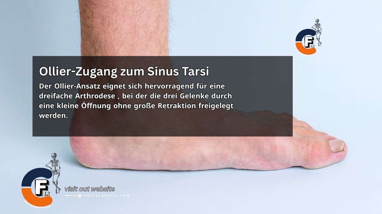
The Ollier approach is an excellent choice for a triple arthrodesis, as it allows access to the three joints through a small opening without the need for extensive retraction. Wound healing is typically good because the proximal flap is created as a full-thickness layer, and the skin edges are protected during retraction.

Patient Positioning

The patient is placed in a supine position with a bump under the ipsilateral buttock.

Landmarks and Incision

  • Make a curved incision, 8–10 cm in length.
  • Begin the incision over the dorsal-lateral aspect of the talonavicular joint.
  • Extend the incision posteriorly across the sinus tarsi (the soft-tissue depression just anterior to the lateral malleolus).
  • Continue the incision obliquely to a point 2.5 cm below the tip of the lateral malleolus.

Internervous Plane

The internervous plane for the Ollier approach lies between:

  • Peroneus tertius muscle: innervated by the deep peroneal nerve.
  • Peroneus brevis muscle: innervated by the superficial peroneal nerve.

Superficial Dissection

  • Incise the fascia and divide the inferior extensor retinaculum along the line of the incision.
  • Ligate any veins crossing the surgical field.
  • Mobilize small skin flaps (large flaps may necrose).
  • Incise the deep fascia and extensor retinaculum in line with the skin incision. Take care to avoid damaging the peroneus tertius and extensor digitorum longus muscles.
  • In the upper (distal) part of the incision, expose the peroneus tertius and extensor digitorum longus and retract them medially.
  • In the lower part of the incision, expose the peroneal tendons and retract them inferiorly.

Deep Dissection

  • Partially resect the fat pad overlying the sinus tarsi using sharp dissection (leave it attached to the skin flap).
  • Identify and release the origin of the extensor digitorum brevis muscle beneath the fat pad.
  • Retract the extensor digitorum brevis muscle distally.
  • Identify and incise the dorsal capsule of the talocalcaneonavicular joint.
  • Identify and incise the capsule of the calcaneocuboid joint.
  • Incise the peroneal retinacula and retract the peroneal tendons anteriorly.
  • Identify and incise the capsule of the posterior talocalcaneal joint.

Dangers

Structures at risk during the Ollier approach include:

  1. Skin flap necrosis: Keep flaps small and full-thickness, and avoid sharp curves.
  2. Tendon of the extensor digitorum longus muscle.
  3. Peroneus brevis tendon.

References & More

  • Campbell’s Operative Orthopaedics, 12th edition.
  • Ollier’s lateral approach to the hindfoot – Orthobullets
  • Meng Q, Wang Q, Wu X, Peng A, Yan J. Clinical application of the sinus tarsi approach in the treatment of intra-articular calcaneal fractures. Medicine (Baltimore). 2018 Mar;97(13):e0175. doi: 10.1097/MD.0000000000010175. PMID: 29595648; PMCID: PMC5895366. Pubmed

Still to read...