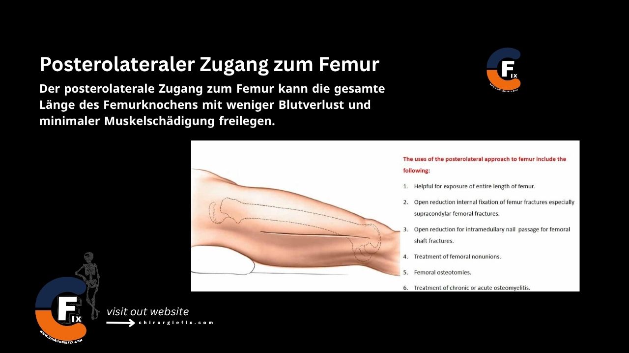
Posterolateral approach to the femur

Posterolateral Approach to the Femur

The posterolateral approach to the femur provides access to the entire length of the femoral shaft while offering the benefits of reduced blood loss and minimal muscle damage.

Common Indications for the Posterolateral Approach

This versatile surgical approach is used for several procedures, including:

  1. Exposing the full length of the femur.
  2. Open internal fixation of femoral fractures, especially supracondylar fractures.
  3. Open reduction to facilitate intramedullary nailing for femoral shaft fractures.
  4. Treatment of femoral nonunions.
  5. Femoral osteotomies.
  6. Management of acute or chronic osteomyelitis.
  7. Biopsy and treatment of bone tumors.

While other lateral approaches involve splitting the vastus lateralis or vastus intermedius muscles, the functional outcomes of the posterolateral approach are comparable. This is likely because the vastus lateralis muscle partially originates from the lateral intermuscular septum.

Patient Positioning

Place the patient supine on the operating table. Position a sandbag under the affected side of the buttock to elevate it and allow the leg to internally rotate. This maneuver lifts the posterolateral thigh surface away from the table.

Landmarks and Incision

  • Landmarks: The lateral femoral epicondyle is the key landmark.
  • Incision: Make a longitudinal incision on the posterolateral aspect of the thigh. Begin the distal portion of the incision at the lateral femoral epicondyle and extend it proximally along the posterior border of the femoral shaft. The total length of the incision depends on the specific procedure being performed.

Internervous Plane

The internervous plane for the posterolateral approach lies between the following structures:

  • Vastus lateralis muscle: Innervated by the femoral nerve.
  • Lateral intermuscular septum: This septum covers the thigh muscles that are innervated by the sciatic nerve.

Superficial Dissection

Incise the tensor fascia lata in line with its fibers, following the skin incision.

Deep Dissection

  • Locate the vastus lateralis muscle beneath the fascia lata.
  • Trace the muscle posteriorly to the lateral intermuscular septum. Reflect the muscle anteriorly and dissect the plane between the muscle and the septum.
  • Begin at the distal end of the incision, where this plane is easiest to identify and develop. Numerous perforating artery branches cross this septum to supply the muscle; these must be ligated or coagulated.
  • If the approach involves the supracondylar area, identify and ligate the numerous branches of the superior lateral genicular artery that cross the operative field. Failure to do so can lead to significant bleeding that is difficult to control.
  • Continue the dissection along the plane between the lateral intermuscular septum and the vastus lateralis muscle. Detach the portions of the vastus lateralis arising from the septum until you reach the femur at the linea aspera.
  • Incise the periosteum longitudinally at this point. Use subperiosteal dissection to remove the muscles covering the femur. Sharp dissection is typically required to detach muscles from the linea aspera itself.
  • In the distal third of the femur, the plane between the vastus lateralis and the lateral intermuscular septum is easily opened. As you move proximally, the muscle becomes thicker, making it more difficult to lift the muscle mass anteriorly to expose the shaft. To assist, place a Hohmann or Bennett retractor over the anterior aspect of the shaft to lift the vastus lateralis forward.
  • Using a retractor on the lateral intermuscular septum can help open the interval and facilitate the proximal dissection.

Approach Extension

Proximal Extension:

  • The posterolateral approach can be extended proximally to the greater trochanter, allowing exposure of nearly the entire femoral shaft.
  • Be aware that the gluteus maximus tendon lies posterior to the lateral intermuscular septum in the proximal extension.

Distal Extension:

  • The posterolateral approach can be extended distally into a lateral parapatellar approach to the knee joint.
  • This provides excellent visualization of the entire distal femur.
  • This extension is particularly useful for the reduction and fixation of intra-articular fractures of the distal femur.

Dangers

The following structures are at risk during the posterolateral approach to the femur:

  1. Perforating branches of the profunda femoris artery:
    • These branches are at risk as they pierce the lateral intermuscular septum.
    • They should be ligated to prevent hematoma formation.
  2. Superior lateral genicular vessels:
    • These vessels are at risk in the distal area near the femoral condyles.
    • They should be ligated to prevent hematoma formation.

References

  • Book “Surgical Exposures in Orthopaedics” – 4th Edition

Still to read...