Anterolateral approach to the ankle joint

Introduction

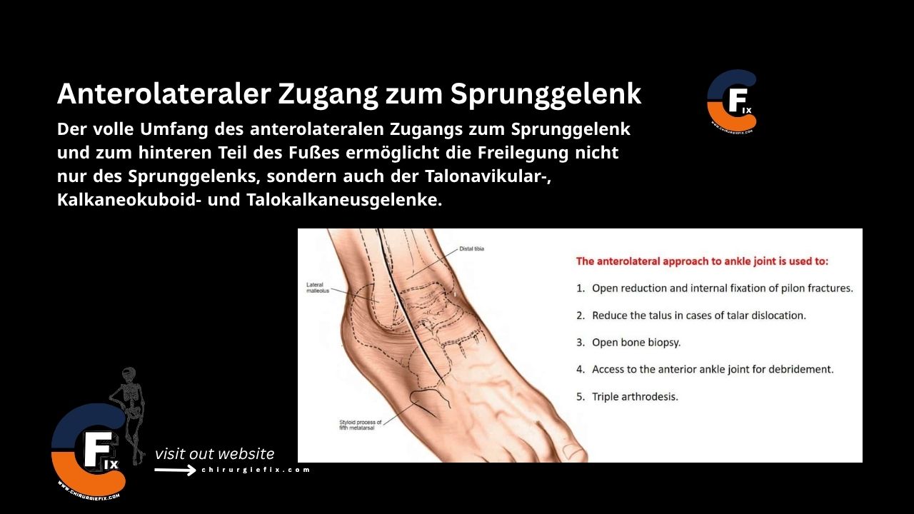
The full anterolateral approach to the ankle joint and hindfoot provides exposure not only of the ankle joint itself but also of the talonavicular, calcaneocuboid, and talocalcaneal joints.

Indications

The anterolateral approach to the ankle is used for:

  1. Open reduction and internal fixation of pilon fractures
  2. Reduction of a dislocated talus
  3. Open bone biopsy
  4. Access to the anterior ankle joint for debridement
  5. Triple arthrodesis

Patient Positioning

Place the patient in a supine position on the operating table. Position a large sandbag under the ipsilateral buttock to internally rotate the leg and bring the lateral malleolus forward.

Landmarks and Incision

Landmarks

  1. Lateral malleolus
  2. Base of the fifth metatarsal

Incision

  • Make a 15 cm long, slightly curved incision on the anterolateral aspect of the ankle.
  • Begin approximately 5 cm proximal to the ankle joint, 2 cm anterior to the anterior border of the fibula.
  • Curve the incision distally, crossing the ankle joint 2 cm medial to the tip of the lateral malleolus, and continue onto the foot. It ends approximately 2 cm medial to the base of the fifth metatarsal, overlying the base of the fourth metatarsal.

Internervous Plane

The internervous plane for the anterolateral approach to the ankle lies between:

  1. The peroneal muscles, supplied by the superficial peroneal nerve.
  2. The extensor muscles, supplied by the deep peroneal nerve.

Superficial Dissection

  • Incise the fascia along the skin incision, cutting through the superior and inferior extensor retinacula.
  • Do not raise skin flaps. Carefully identify and preserve all dorsal cutaneous branches of the superficial peroneal nerve that may cross the dissection field.
  • Identify the peroneus tertius and extensor digitorum longus muscles. In the upper half of the wound, incise directly lateral to these muscles down to the bone.

Deep Dissection

  • Retract the extensor muscles medially to expose the anterior aspect of the distal tibia and the anterior ankle joint capsule.
  • Distally, identify the extensor digitorum brevis muscle at its origin from the calcaneus and release it using sharp dissection.
  • During dissection, branches of the lateral tarsal artery will be encountered; cauterize (diathermy) these to prevent postoperative hematoma formation.
  • Reflect the released extensor digitorum brevis muscle distally and medially, elevating the muscle fascia along with the subcutaneous fat and skin as a single flap.
  • Identify the dorsal capsules of the calcaneocuboid and talonavicular joints, which lie adjacent to each other on the foot and form the clinical midtarsal joint.
  • Next, identify the fat in the sinus tarsi and remove it to expose the talocalcaneal joint. This can be done by either mobilizing the fat pad and reflecting it inferiorly, or by excising it.
  • Preserving the fat pad helps prevent a cosmetically undesirable depression postoperatively. Conserving the wound dressing also supports wound healing.
  • Finally, incise some or all of the exposed capsules. To open the joints, forcefully flex and plantarflex the foot.

Approach Extension

Proximal Extension

The anterolateral approach to the ankle can be extended proximally to explore structures in the anterior compartment of the leg. Continue the incision proximally over the compartment and incise the thick deep fascia along the skin incision.

Distal Extension

The anterolateral approach to the ankle can also be extended distally to expose the tarsometatarsal joints on the lateral aspect of the foot. Continue the incision over the fourth metatarsal and expose the subcutaneous tarsometatarsal joints.

Dangers

Structures at risk during the anterolateral approach to the ankle include:

  1. Superficial peroneal nerve
  2. Deep peroneal nerve
  3. Anterior tibial artery

References

  • Surgical Exposures in Orthopaedics – 4th Edition
  • Campbell’s Operative Orthopaedics, 12th Edition
  • Youtube

Still to read...