Introduction to the Medial Approach to the Elbow
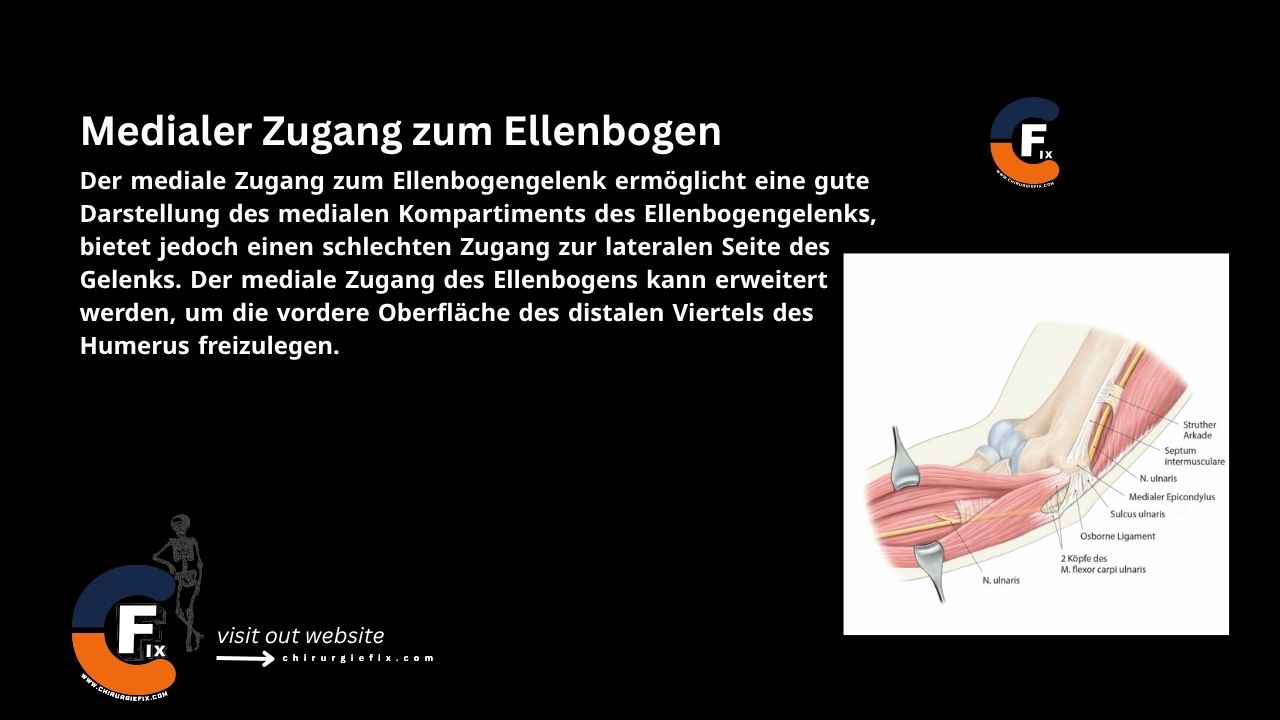
The medial approach to the elbow joint provides excellent visualization of the medial compartment but offers limited access to the lateral side. This approach can be extended to expose the anterior surface of the distal quarter of the humerus.
Indications for the Medial Elbow Approach
- Decompression and/or transposition of the ulnar nerve.
- Removal of loose bodies from the ulnar side.
- Open reduction and internal fixation (ORIF) of the coronoid process of the ulna.
- Open reduction and internal fixation of the medial humeral condyle and epicondyle.
- Debridement and reattachment of the common flexor tendon for medial epicondylitis.
This approach is not suitable for routine elbow examination due to poor lateral access. However, the joint can be dislocated during the procedure to gain access to the lateral side if necessary.
Patient Positioning
Place the patient in the supine position on the operating table. The arm should be flexed and supported by an arm board positioned over the patient. Abduct the arm and externally rotate the shoulder fully so the medial epicondyle of the humerus faces anteriorly. Flex the elbow to 90 degrees.
Alternatively, flex the patient’s shoulder and elbow so the forearm rests across the front of the face. This positioning allows easier access to the medial elbow but requires an assistant to hold the forearm securely for adequate exposure.
Landmarks and Incision
Landmarks
- Medial epicondyle of the humerus.
Incision
Make a curved incision 8 to 10 cm long on the medial side of the elbow, centering it over the medial epicondyle.
Internervous Plane
Proximal Internervous Plane
- Brachialis muscle: innervated by the musculocutaneous nerve.
- Triceps muscle: innervated by the radial nerve.
Distal Internervous Plane
- Brachialis muscle: innervated by the musculocutaneous nerve.
- Pronator teres muscle: innervated by the median nerve.
Superficial Dissection
Incise the fascia over the ulnar nerve, starting proximally, and isolate the nerve along the entire length of the incision. Expose the origin of the common flexor tendon at the medial epicondyle. Develop the interval between the brachialis and pronator teres muscles. Avoid the median nerve, which enters the pronator teres near the midline.
If necessary, perform an osteotomy of the medial epicondyle, reflecting it distally. Ensure the medial collateral ligament remains attached to the osteotomy fragment.
Develop the interval between the brachialis and triceps muscles.
Deep Dissection
Incise the capsule and the medial collateral ligament to expose the joint.
Approach Extension
The medial approach to the elbow can be extended proximally and distally as follows:
Local Extension
- Abducting the forearm opens the medial part of the joint.
- Detaching the joint capsule and periosteum allows lateral dislocation of the joint.
Proximal Extension
- The anterior surface of the distal quarter of the humerus can be exposed by developing the plane between the brachialis and triceps muscles.
- Subperiosteal dissection and elevation of the brachialis muscle expose the anterior surface of the distal quarter of the humerus.
Distal Extension
- The medial epicondyle of the humerus with its attached flexor muscles can only be retracted as far as the branches of the median nerve allow.
- This exposure provides an adequate view of the brachialis insertion on the coronoid but does not allow further distal exposure of the ulna.
Dangers
Structures at risk during the medial approach to the elbow include:
- Ulnar nerve: At risk, must be identified and protected throughout the approach.
- Median nerve: Aggressive retraction of the osteotomy fragment can cause traction injury to the median nerve and the anterior interosseous nerve.
References
- Book: “Surgical Exposures in Orthopaedics” – 4th Edition
- Campbell’s “Operative Orthopaedics”, 12th Edition
- Orthobullets Website