Posterior approach to the elbow

The posterior approach to the elbow provides the best possible view of the bones that make up the elbow joint. This approach typically requires an osteotomy of the olecranon at its articular surface, creating an additional “fracture” that must be internally fixed.

Indications for the Posterior Approach to the Elbow

  1. Open reduction and internal fixation of distal humerus fractures: This approach offers the best possible intra-articular view of the elbow joint.
  2. Removal of loose bodies within the joint.
  3. Treatment of distal humerus nonunions.
  4. Triceps muscle lengthening for elbow extension contractures.

Patient Positioning

  • Position the patient prone or in the lateral decubitus position with the elbow flexed and the arm hanging over the side of the table.

Landmarks and Incision

  • Landmarks:
    • Olecranon process.
  • Incision:
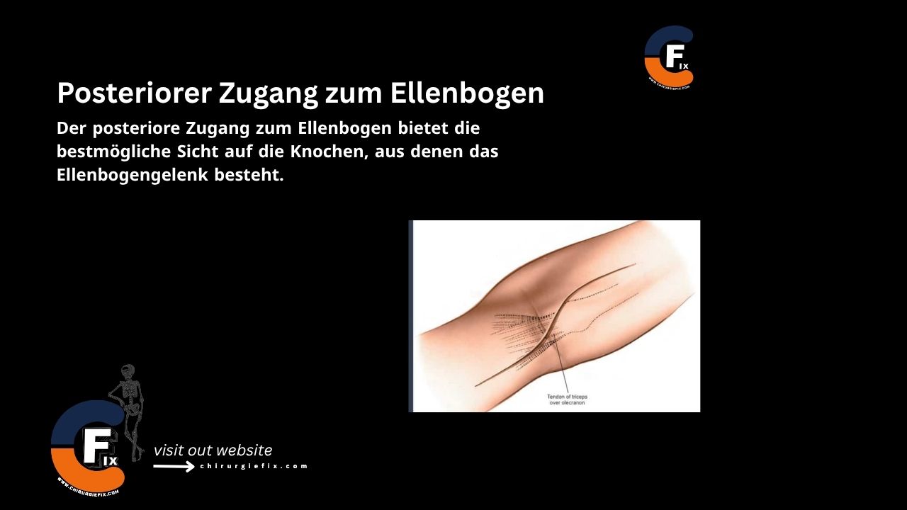
    • Make a longitudinal incision on the posterior aspect of the elbow. Begin 5 cm above the olecranon in the midline of the posterior arm.
    • Just proximal to the tip of the olecranon, curve the incision laterally so that it runs along the lateral side of the process.
    • To complete the incision, curve it back medially so that it lies over the center of the subcutaneous surface of the ulna.
    • By curving the incision around the tip of the olecranon, the suture line is moved away from the implants used to fix the olecranon osteotomy and away from the weight-bearing tip of the elbow.

Internervous Plane

There is no true internervous plane for the posterior approach to the elbow:

  • The extensor mechanism (the triceps muscle) is either split or detached.
  • The radial nerve innervates the triceps muscle further proximally.

Superficial Dissection

  • First, palpate the ulnar nerve and fully dissect it out:
    • It is helpful to place a vessel loop or Penrose drain around it for identification at all times.
  • Incise the deep posterior fascia in the midline.
  • You can either split the triceps fascia or proceed with the olecranon osteotomy.
  • If performing an olecranon osteotomy, drill and tap the olecranon before performing the osteotomy.
  • Score the olecranon with an osteotome to allow for perfect repositioning during osteotomy repair.
  • Perform a V-shaped osteotomy of the olecranon 2 cm from its tip using an oscillating saw.

Deep Dissection

  • Remove soft tissue from the edges of the osteotomy site and retract the olecranon fragment proximally.
  • Subperiosteal dissection of the medial and lateral borders of the humerus allows exposure of the entire distal quarter of the humerus.

Extension of the Approach

The posterior approach to the elbow can be extended as follows:

  • Proximal extension:
    • The posterior approach to the elbow cannot be extended proximally beyond the distal third of the humerus due to the risk to the radial nerve.
  • Distal extension:
    • The incision can be continued along the subcutaneous border of the ulna, exposing the entire length of this bone.

Dangers

Structures at risk during the posterior approach to the elbow include:

  1. Ulnar nerve:
    • The ulnar nerve should be identified and protected first in this approach.
    • It can usually be palpated 2 cm proximal to the medial epicondyle.
    • Transposition of the ulnar nerve has not been shown to reduce the incidence of ulnar neuritis.
  2. Median nerve:
    • Strict subperiosteal dissection of the anterior surface of the humerus protects the nerve.
    • Flexing the elbow relaxes the anterior structures.
  3. Radial nerve:
    • The radial nerve is at risk proximally as it travels from the posterior to the anterior brachial compartment through the lateral intermuscular septum.
    • It is typically located on the lateral border of the humerus near the distal third junction.
  4. Brachial artery:
    • The brachial artery travels with the median nerve.

References

  • Book “Surgical Exposures in Orthopaedics” – 4th Edition
  • Campbell’s “Operative Orthopaedics”, 12th Edition

Still to read...