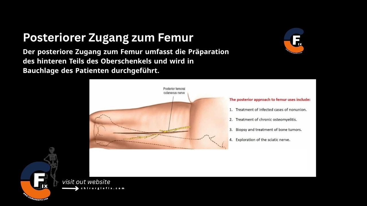
Posterior approach to the femur

The posterior approach to the femur involves dissecting the back of the thigh and is performed with the patient in the prone position. The key to this approach lies in understanding the anatomy of the sciatic nerve and its relationship to the biceps femoris muscle.

The posterior approach to the femur is used for:

  1. Treating infected cases of nonunion.
  2. Managing chronic osteomyelitis.
  3. Biopsy and treatment of bone tumors.
  4. Exploration of the sciatic nerve.

Patient Positioning

Place the patient in the prone position on the operating table.

Landmarks and Incision

Landmarks:

  • The gluteal fold.

Incision:

  • Make a straight longitudinal incision approximately 20 cm long along the midline of the posterior thigh.
  • The incision should end proximally at the lower edge of the gluteal fold; its length varies according to surgical needs.

Internervous Plane

The internervous plane for the posterior approach to the femur lies between:

  • The lateral intermuscular septum, which covers the vastus lateralis muscle (innervated by the femoral nerve).
  • The biceps femoris muscle (innervated by the sciatic nerve).

Superficial Dissection

  • Incise the deep fascia of the thigh along or just lateral to the skin incision, being careful to avoid damaging the posterior femoral cutaneous nerve. This nerve runs longitudinally under the deep fascia (and roughly in line with the fascial incision) in the groove between the biceps femoris and semitendinosus muscles.
  • Identify the lateral border of the biceps femoris at the proximal end of the wound by palpation.
  • Develop the plane between the biceps femoris and the vastus lateralis muscles, which is covered by the lateral intermuscular septum.

Deep Dissection

  • Begin proximally by retracting the long head of the biceps femoris medially and the lateral intermuscular septum laterally, developing the plane with a finger.
  • Identify the short head of the biceps, which originates from the lateral lip of the linea aspera.
  • Detach its origin from the femur using sharp dissection and reflect it medially to expose the posterior surface of the femur.
  • In the distal half of the wound, retract the long head of the biceps laterally to expose the sciatic nerve. Note that the nerve may have already divided into its tibial and common peroneal branches; in this case, two "sciatic nerves" run side by side.
  • Gently retract the sciatic nerve laterally to expose the posterior part of the femur, which is covered by periosteum.
  • Develop an epiperiosteal plane between the periosteum and the overlying soft tissues.

Approach Extension

  • The posterior approach to the femur cannot be meaningfully extended proximally or distally.
  • Its value lies in providing exposure to the middle three-fifths of the femoral shaft.

Dangers

Structures at risk during the posterior approach to the femur include:

  1. Posterior femoral cutaneous nerve.
  2. Sciatic nerve.
  3. Nerve to the biceps femoris.

Semimembranosus Muscle

  • The attachments of the semimembranosus muscle significantly reinforce the posterior and posteromedial joint capsule of the knee.
  • The muscle can be transferred, along with the semitendinosus tendon, to the anterior surface of the lateral femoral condyle to correct an internal rotation deformity of the hip in patients with various neurological lesions, a technique that is rarely used.

Semitendinosus Muscle

  • As its name suggests, the semitendinosus muscle has an extremely long tendon relative to its muscle belly. The tendon is at least 13 cm long and can be used in various surgical procedures.
  • It can be left attached to the tibia even when secured through a drill hole in the patella to hold this bone medially in cases of recurrent dislocation.
  • It can also be used for reconstruction of the posterior and anterior cruciate ligaments. In this procedure, the tendon is detached from the muscle at the musculotendinous junction and passed through the femur to mimic the function of the missing cruciate ligaments. Additionally, it can be used to reinforce a torn medial collateral ligament in the knee.

References

  • Book "Surgical Exposures in Orthopaedics" – 4th Edition
  • Campbell's "Operative Orthopaedics", 12th Edition
  • BOSWORTH DM: Posterior approach to the femur. J Bone Joint Surg 26:687, 1944.

Still to read...