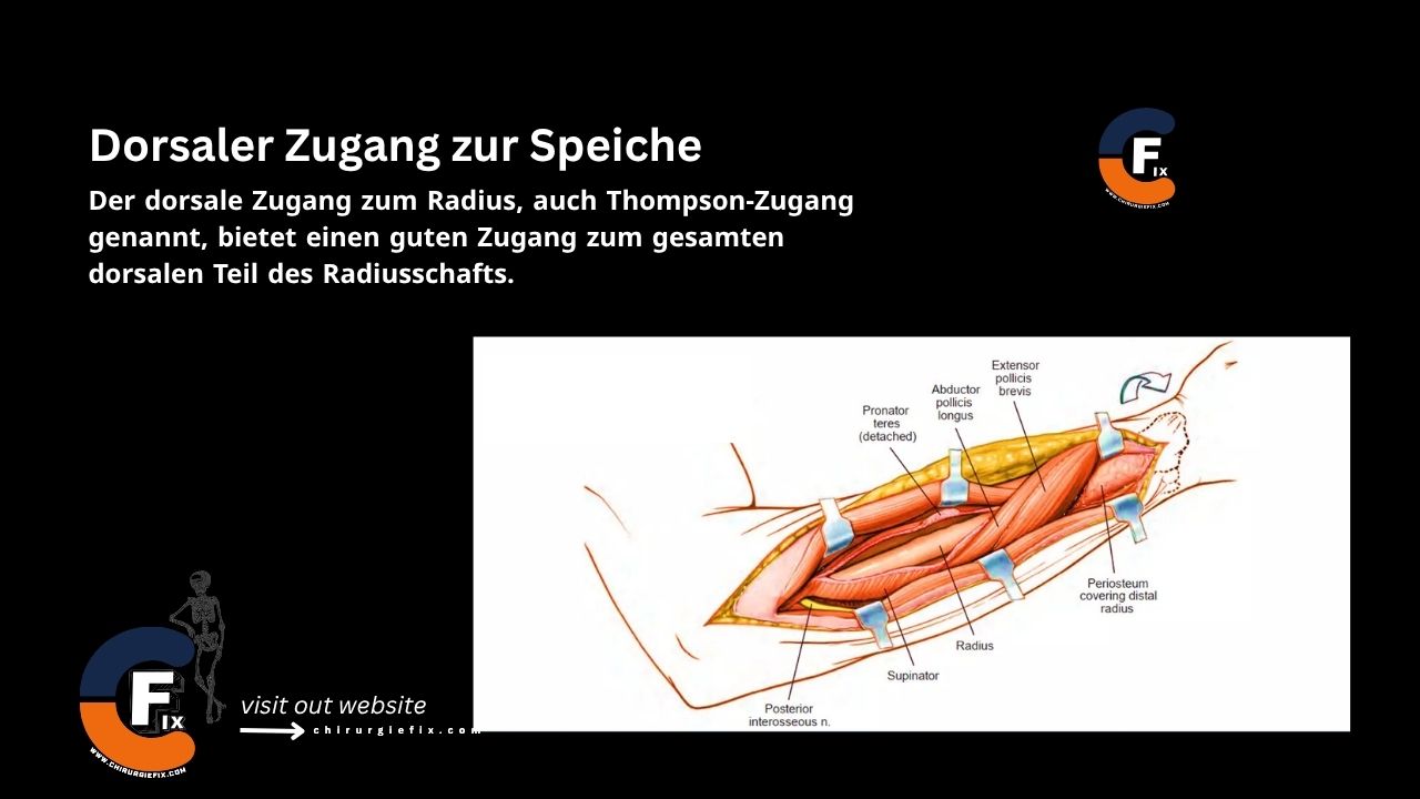
The dorsal approach to the radius, also known as the Thompson approach, provides excellent exposure to the entire dorsal aspect of the radial shaft.
The primary goal of this approach is to isolate and retract the posterior interosseous nerve before exposing the most proximal parts of the radial shaft. This keeps the nerve under direct observation throughout the subsequent procedure, protecting it from injury.
Uses of the posterior approach to the radius include:
Place the patient in one of two positions:
Make a straight incision running from a point just anterior to the lateral epicondyle of the humerus (along the back of the forearm) to a point just distal to the ulnar side of Lister's tubercle at the wrist.
Usually, only a portion of this incision is needed for surgery. In the case of a fracture, the incision should be centered over the fracture site. Using an image intensifier can allow for more accurate placement of the incision.
Proximal: The internervous plane for the dorsal approach to the radius lies between the extensor carpi radialis brevis (supplied by the radial nerve) and the extensor digitorum communis (supplied by the posterior interosseous nerve). The common aponeurosis of these muscles is the splitting plane.
Distal: The internervous plane for the dorsal approach to the radius lies between the extensor carpi radialis brevis (supplied by the radial nerve) and the extensor pollicis longus (supplied by the posterior interosseous nerve).
Incise the deep fascia along the skin incision and identify the space between the extensor carpi radialis brevis and the extensor digitorum communis.
Distally, this plane is more apparent, where the abductor pollicis longus and extensor pollicis brevis emerge between the two muscles. Proximally, the extensor carpi radialis brevis and extensor digitorum communis share a common aponeurosis. Continue the dissection proximally, separating the two muscles to expose the upper third of the radial shaft, which is covered by the enveloping supinator muscle.
Below the abductor pollicis longus and extensor pollicis brevis, identify the intermuscular plane between the extensor carpi radialis brevis and extensor pollicis longus. Separating these two muscles exposes the lateral part of the radial shaft.
The supinator muscle conceals the dorsal side of the upper third of the radius; the posterior interosseous nerve runs within its substance between the superficial and deep heads. The nerve emerges between the superficial and deep heads of the supinator muscle about 1 cm proximal to the distal edge of the muscle. At this point, it divides into branches that supply the extensors of the wrist, fingers, and thumb.
There are two methods to successfully identify and preserve this nerve during its course through the muscle:
Once the nerve has been successfully identified and preserved, fully supinate the arm to visualize the anterior surface of the radius. Release the attachment of the supinator muscle from the anterior side of the radius. Subperiosteally elevate the supinator off the bone to expose the proximal third of the radial shaft.
Two muscles, the abductor pollicis longus and extensor pollicis brevis, cover this approach as they cross the dorsal side of the radius before passing distally and radially over the middle third of the radius. To remove them from the bone, make an incision along their upper and lower borders. They can then be easily separated from the underlying radius and retracted either distally or proximally, depending on the exposure required. Plates can be slid under these muscles for fixation if needed.
The separation of the extensor carpi radialis brevis from the extensor pollicis longus has already led directly to the lateral border of the radius.
The Thompson approach can be extended to the dorsum of the wrist (see Dorsal Approach to the Wrist).
The Thompson approach can be extended proximally to expose the lateral epicondyle of the humerus (see Lateral Approach to the Distal Humerus). However, these extensions are rarely necessary.
There are two ways to preserve the critical posterior interosseous nerve during the dorsal approach to the radius, which is the key to this dissection: