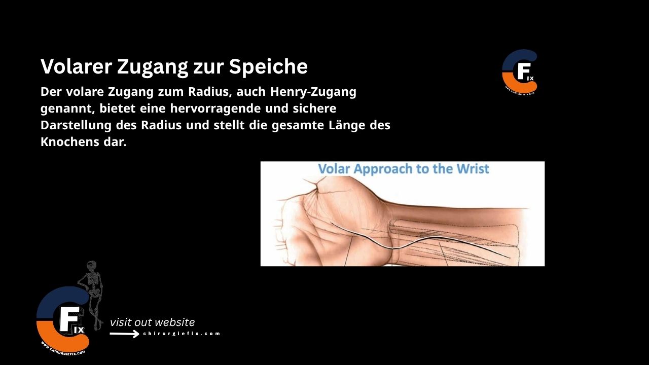
The volar approach to the radius, also known as the Henry approach, provides excellent and safe exposure of the radius, revealing the full length of the bone.
The volar approach to the radius is used for:
Place the patient supine with the arm extended on an arm board. Fully supinate the forearm.
Make a straight incision from the anterior elbow crease, just lateral to the biceps tendon, extending distally to the radial styloid process.
The internervous plane for the Henry approach is as follows:
Incise the deep fascia along the skin incision. Develop the plane between the brachioradialis and flexor carpi radialis distally. Moving proximally, develop the plane between the pronator teres and brachioradialis. Identify the superficial radial nerve beneath the brachioradialis muscle. Ligate the branches of the radial artery to facilitate lateral retraction of the brachioradialis.
Follow the biceps tendon to its insertion on the bicipital tuberosity. Incise the bursa radially to the biceps tendon insertion to access the proximal radius. The radial artery runs along the ulnar side of the biceps tendon. Fully supinate the forearm to move the posterior interosseous nerve radially and bring the origin of the supinator muscle to the anterior aspect of the radius. Incise the supinator along the line of its broad insertion and continue subperiosteal dissection laterally.
Pronate the forearm to visualize the pronator teres insertion along the radial aspect of the radius. Release the pronator teres insertion from the bone and retract it medially.
Partially supinate the forearm and elevate the periosteum from the lateral side of the distal third of the radius, lateral to the pronator quadratus and flexor pollicis longus muscles.
The volar approach to the radius provides complete access to the entire length of the bone. It can be extended distally to expose the wrist joint.
Structures at risk during the volar approach to the radius (Henry approach) include:
The posterior interosseous nerve enters the supinator muscle under a fibrous arch known as the arcade of Frohse. This arch is formed by the thickened edge of the superficial head of the supinator. Compression of the nerve at this point can lead to paralysis or dysfunction of the extensor muscles, known as posterior interosseous nerve entrapment syndrome.
Measures to protect the posterior interosseous nerve include:
Injury results in neurapraxia, which may take 6–9 months to heal.
The superficial radial nerve courses along the forearm beneath the brachioradialis muscle. It is at risk during manipulation of the mobile wad. Damage can result in a painful neuroma.
The radial artery runs in the mid-forearm beneath the brachioradialis muscle. The radial recurrent arteries are a series of vessels arising from the radial artery just below the elbow joint. They consist of anterior and posterior groups that pass anterior and posterior to the superficial radial nerve before entering the brachioradialis. These vessels must be ligated to allow mobilization of both the artery and nerve.