Hardinge Access

Hardinge Access

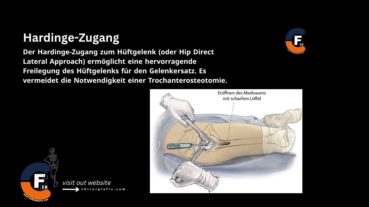
The Hardinge approach to the hip joint (also known as the direct lateral approach) provides excellent exposure for hip replacement surgery. This technique eliminates the need for a trochanteric osteotomy.

It is important to note that this approach offers less extensive exposure than the anterolateral approach, which does require a trochanteric osteotomy.

Indications for the Hardinge Approach

The Hardinge approach is used for:

  1. Total Hip Arthroplasty: This approach is associated with a lower rate of hip prosthesis dislocation.
  2. Proximal Femur Fractures.

Patient Positioning

  1. Lateral Position.
  2. Supine Position: The patient is placed supine with the greater trochanter positioned at the edge of the table. This allows the gluteal muscles and fat to fall posteriorly, away from the surgical field.

Landmarks and Incision

  • Landmarks:
    1. Anterior superior iliac spine.
    2. Lateral aspect of the greater trochanter.
    3. Shaft of the femur.
  • Incision:
    • Begin the incision 5 cm proximal to the tip of the greater trochanter.
    • Make a longitudinal incision that passes over the center of the greater trochanter’s tip and extends approximately 8 cm along the femoral shaft.

Internervous Plane

There is no true internervous plane for the Hardinge approach. The fibers of the gluteus medius muscle are split in line, distal to the point where the superior gluteal nerve supplies the muscle. The vastus lateralis muscle is also split in line, lateral to the point where it is innervated by the femoral nerve.

Superficial Dissection

  • Incise the subcutaneous fat and the underlying deep fascia along the line of the skin incision.
  • Retract the fascial edges, drawing the tensor fasciae latae anteriorly and the gluteus maximus posteriorly.
  • Sharply dissect any gluteus medius fibers that adhere to the deep surface of the fascia. The vastus lateralis and gluteus medius are now exposed.

Deep Dissection

  • Split the fibers of the gluteus medius in line with their direction, starting at the midpoint of the greater trochanter. Do not extend the split more than 3 cm proximal to the trochanter’s edge, as this could damage branches of the superior gluteal nerve.
  • Divide the fibers of the vastus lateralis that lie over the lateral side of the base of the greater trochanter.
  • Develop an anterior flap consisting of the anterior part of the gluteus medius, the underlying gluteus minimus, and the anterior part of the vastus lateralis.
  • Detach the muscles from the greater trochanter, using either sharp dissection or by raising a small bone flake.
  • Continue developing this anterior flap, following the contour of the bone onto the femoral neck, until the anterior hip joint capsule is fully exposed.
  • Release the attachment of the gluteus minimus tendon from the anterior part of the greater trochanter.
  • Develop the plane between the hip joint capsule and the overlying muscles by bluntly inserting a swab into the potential space.
  • Open the capsule with a T-shaped incision.
  • Osteotomize the femoral neck and remove the femoral head using a corkscrew.
  • Complete the exposure of the acetabulum by inserting appropriate retractors around it.

Approach Extension

Distal Extension:

The Hardinge approach can be easily extended distally. To expose the femoral shaft, split the vastus lateralis muscle in line with its fibers (as in a lateral approach to the femur).

Proximal Extension:

The Hardinge approach cannot be extended proximally.

Dangers

Structures at risk during the Hardinge approach include:

  1. Superior Gluteal Nerve:
    • The superior gluteal nerve runs between the gluteus medius and gluteus minimus muscles, 3–5 cm proximal to the greater trochanter.
    • It can be protected by limiting the proximal incision into the gluteus medius and placing a stay suture at the apex of the gluteal split.
    • Injury to this nerve can lead to a Trendelenburg gait.
  2. Femoral Nerve:
    • The femoral nerve is the most lateral structure in the anterior thigh’s neurovascular bundle.
    • Always keep retractors on bone and avoid placing them over soft tissue to prevent iatrogenic injury.

References

  1. Book: “Surgical Exposures in Orthopaedics” – 4th Edition
  2. Campbell’s “Operative Orthopaedics”, 12th Edition
  3. Hardinge K. The direct lateral approach to the hip. J Bone Joint Surg Br 1982;64B:17–18. Link

Still to read...