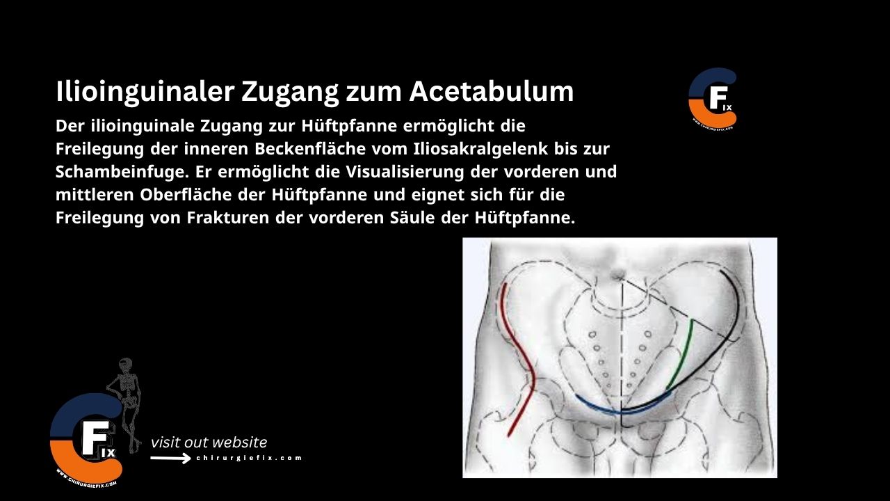
Ilioinguinal approach to the acetabulum

Introduction

The ilioinguinal approach to the acetabulum provides exposure of the inner pelvic surface from the sacroiliac joint to the pubic symphysis. It allows visualization of the anterior and middle surfaces of the acetabulum and is suitable for exposing fractures of the anterior column of the acetabulum.

Indications for the Ilioinguinal Approach

This approach is used for the following fracture types:

  1. Anterior wall fractures
  2. Anterior column fractures
  3. Anterior column plus posterior hemitransverse fractures
  4. Open reduction and internal fixation of most both-column fractures
  5. Any T-type fracture
  6. Some transverse fractures
  7. Transverse fractures with anterior displacement

Patient Positioning

  • Place the patient supine on the operating table, with the greater trochanter on the fracture side positioned at the edge of the table.
  • Place a bump under the ipsilateral buttock.
  • Flex the affected leg to relax the iliopsoas and neurovascular structures.

Landmarks and Incision

Landmarks

  1. The anterior superior iliac spine (ASIS)
  2. The pubic tubercle

Incision

  • Make a curved anterior incision starting 5 cm above the anterior superior iliac spine.
  • Extend the incision medially so it runs 1 cm above the pubis and ends at the midline.

Internervous Plane

  • There is no true internervous plane for the ilioinguinal approach to the acetabulum.
  • The dissection essentially involves lifting muscle, nerve, and vascular structures away from the inner wall of the pelvis.

Superficial Dissection

The superficial dissection for the ilioinguinal approach is performed as follows:

  • Dissect through the subcutaneous fat to expose the aponeuroses of the external oblique muscle.
  • The aponeurosis of the external oblique muscle is divided along the direction of its fibers, from the superficial inguinal ring to the anterior superior iliac spine. This exposes the spermatic cord in males and the round ligament in females.
  • Gently isolate these structures in a sling.
  • Continue the dissection medially and divide the anterior part of the rectus sheath to expose the underlying rectus abdominis muscle.
  • Elevate the iliacus muscle from the inner aspect of the iliac wing. Begin with sharp dissection; once inside the pelvis, use blunt dissection.

Deep Dissection

The deep dissection for the ilioinguinal approach is performed as follows:

  • Bluntly dissect a plane between the pubic symphysis and bladder (space of Retzius) and pack it with sponges.
  • Expose the anterior part of the femoral vessels and surrounding lymphatics in the middle of the incision (lacuna vasorum).
    • The lacuna musculorum is located laterally and contains the iliopsoas muscle, femoral nerve, and lateral femoral cutaneous nerve.
  • Identify the iliopectineal fascia, which separates the lacuna vasorum and lacuna musculorum.
  • Dissect the vessels and lymphatics from the medial aspect of the fascia; free the iliopsoas and femoral nerve from the lateral aspect.
  • Sharply divide the iliopectineal fascia down to the iliopectineal eminence, then release it from the pelvic brim. This provides access to the true pelvis, quadrilateral plate, and posterior column.
  • Place a second Penrose drain around the iliopsoas, femoral nerve, and lateral femoral cutaneous nerve.
  • Place a third Penrose drain around the femoral vessels and lymphatics.
  • Identify and ligate the corona mortis before retracting the vessels.
  • Use subperiosteal dissection to expose the pelvic brim, rami, and quadrilateral surface.

Working Through Three Windows to Reduce and Fix the Fracture

  1. Medial window:
    • Medial to the external iliac artery and vein.
    • Provides access to the pubic rami and indirect access to the inner pelvic fossa and anterior sacroiliac joint.
  2. Middle window:
    • Between the external iliac vessels and the iliopsoas muscle.
    • Provides access to the pelvic brim, quadrilateral plate, and part of the superior pubic ramus.
  3. Lateral window:
    • Lateral to the iliopsoas (iliopectineal fascia).
    • Provides access to the quadrilateral plate, sacroiliac joint, and iliac wing.

Approach Extension

Proximal Extension

  • The ilioinguinal approach can be extended proximally to expose the sacroiliac joint.
  • Extend the skin incision posteriorly, following the iliac crest.
  • Use sharp dissection down to the bone.
  • Strip the origins of the iliacus muscle from the inner aspect of the ilium using blunt dissection.
  • Retract the iliacus medially to expose the inner wall of the ilium and the sacroiliac joint.

Distal Extension

  • The ilioinguinal approach cannot be extended distally.

Dangers

Structures at risk during the ilioinguinal approach include:

  1. Femoral nerve.
  2. Femoral and external iliac arteries:
    • Damage can lead to thrombosis.
    • Protect them by keeping them within the femoral sheath.
  3. Lymphatics:
    • Present in the fatty areolar tissue around the vessels.
    • Disruption can impair postoperative lymphatic drainage and cause edema.
  4. Lateral femoral cutaneous nerve: Often must be sacrificed, leading to numbness on the lateral thigh.
  5. Inferior epigastric artery: Must be ligated if it has an anomalous origin from the obturator artery to allow retraction of the pelvic vessels.
  6. Spermatic cord (contains vas deferens and testicular artery):
    • Must be protected.
    • Damage can lead to testicular ischemia and infertility.
  7. Heterotopic ossification: Much more common with the extended iliofemoral and Kocher-Langenbeck approaches.
  8. Obturator nerve: Injury causes numbness in the medial thigh.

References

  • Surgical Exposures in Orthopaedics – 4th Edition
  • Campbell’s Operative Orthopaedics – 12th Edition

Still to read...