Posterior approach to the sacroiliac joint

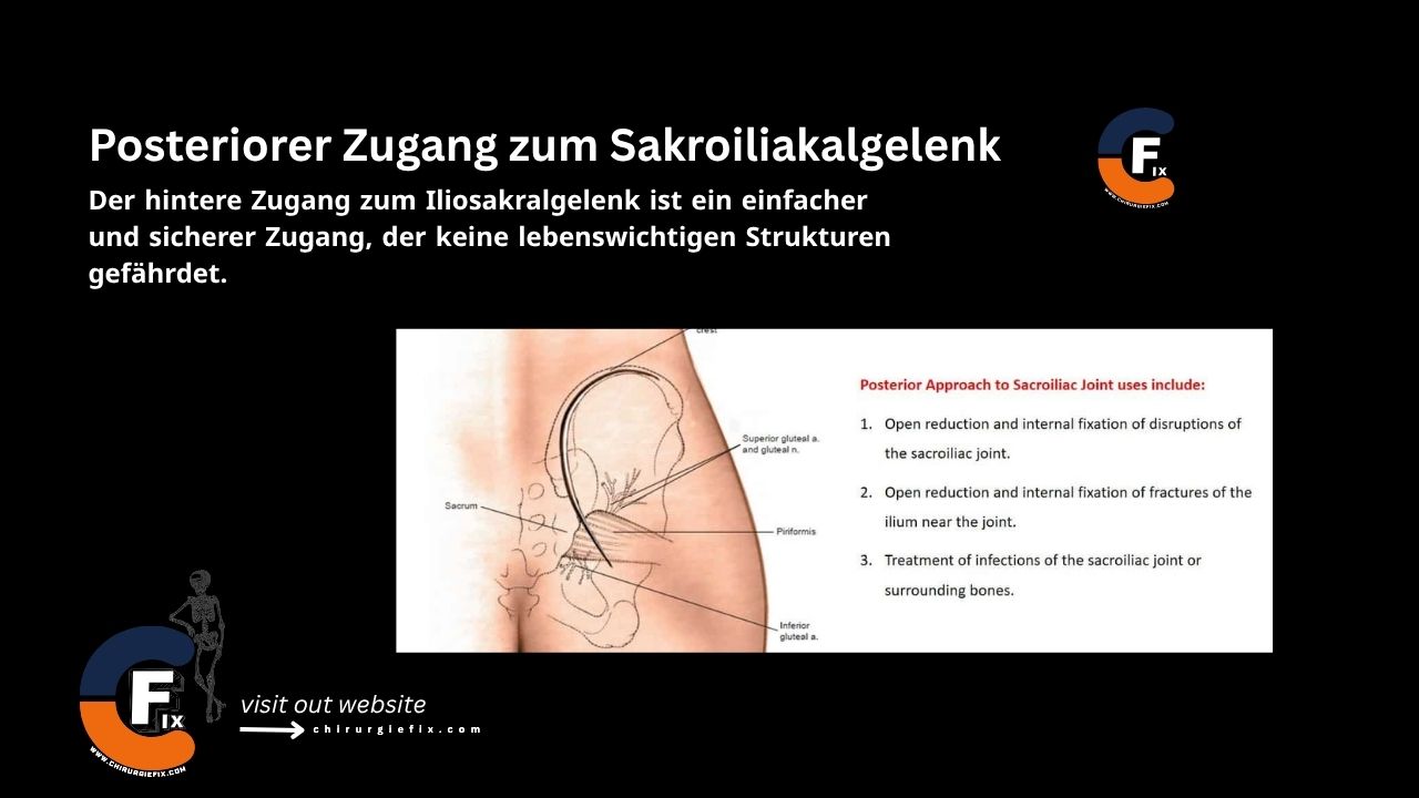
The posterior approach to the sacroiliac joint is a straightforward and safe surgical route that does not endanger vital structures.

Indications for the posterior approach to the sacroiliac joint include:

  1. Open reduction and internal fixation of sacroiliac joint disruptions.
  2. Open reduction and internal fixation of fractures of the iliac bone adjacent to the joint.
  3. Treatment of infections of the sacroiliac joint or surrounding bone.

The popularity of this approach has declined with the increasing use of percutaneous screw fixation techniques.

It is important to note that reduction of fractures and dislocations through this approach is difficult, particularly the correction of vertical displacement.

Patient Positioning

  • Place the patient in a prone position on the operating table.
  • Position the pads longitudinally to support the chest wall and pelvis. The pads should allow expansion of the chest wall and abdomen without contacting the table.

Landmarks and Incision

  • Landmarks:
    1. Posterior iliac crest.
    2. Posterior superior iliac spine.
  • Incision:
    • Make a curved incision over the posterior iliac crest, starting 3 cm distal and lateral to the posterior superior iliac spine.
    • Extend the incision from this point to the posterior superior iliac spine, and then continue it along the crest to its highest point.

Internervous Plane

  • There is no true internervous plane for the posterior approach to the sacroiliac joint.
  • Both the gluteus maximus and gluteus medius must be partially detached from their origins, but their individual neurovascular pedicles can be easily preserved.

Superficial Dissection

  • Divide the subcutaneous tissue along the skin incision.
  • Incise the outer edge of the subcutaneous surface of the iliac crest to expose the fascial layer covering the gluteus maximus muscle.
  • Detach the origin of the gluteus maximus muscle from the crest and gently reflect the muscle downward and laterally.
  • As the gluteus maximus is reflected, the gluteus medius and piriformis muscles are exposed as they emerge through the greater sciatic notch.

Deep Dissection

  • Gently lift the gluteus medius muscle from the outer wing of the ilium.
  • The muscle cannot be lifted far anteriorly because its deep surface is connected by its neurovascular bundle, the superior gluteal nerve and vessels.
  • In trauma, the disruption or fracture of the sacroiliac joint is clearly visible, but reduction is extremely difficult.
  • To assess reduction, detach part of the origin of the piriformis muscle around the greater sciatic notch and insert a finger through the notch to palpate the joint from its anterior surface.
  • The joint surface will feel smooth when it has been reduced.

Approach Extension

  • The posterior approach to the sacroiliac joint can be extended anteriorly by lifting the gluteus medius and gluteus minimus muscles from the outer aspect of the iliac wing.
  • This allows for the treatment of larger fractures of the iliac wing and ilium.

Dangers

Structures at risk during the posterior approach to the sacroiliac joint include:
  • Nerves:
    1. Inferior gluteal nerve.
    2. Superior gluteal nerve.
    3. Sacral nerve roots.
  • Vessels:
    • Branches of the superior and inferior gluteal arteries run with their respective nerves and are also at risk.

References

  • Surgical Exposures in Orthopaedics – 4th Edition
  • Campbell's Operative Orthopaedics, 12th Edition

Still to read...