Anterior approach to the tibia

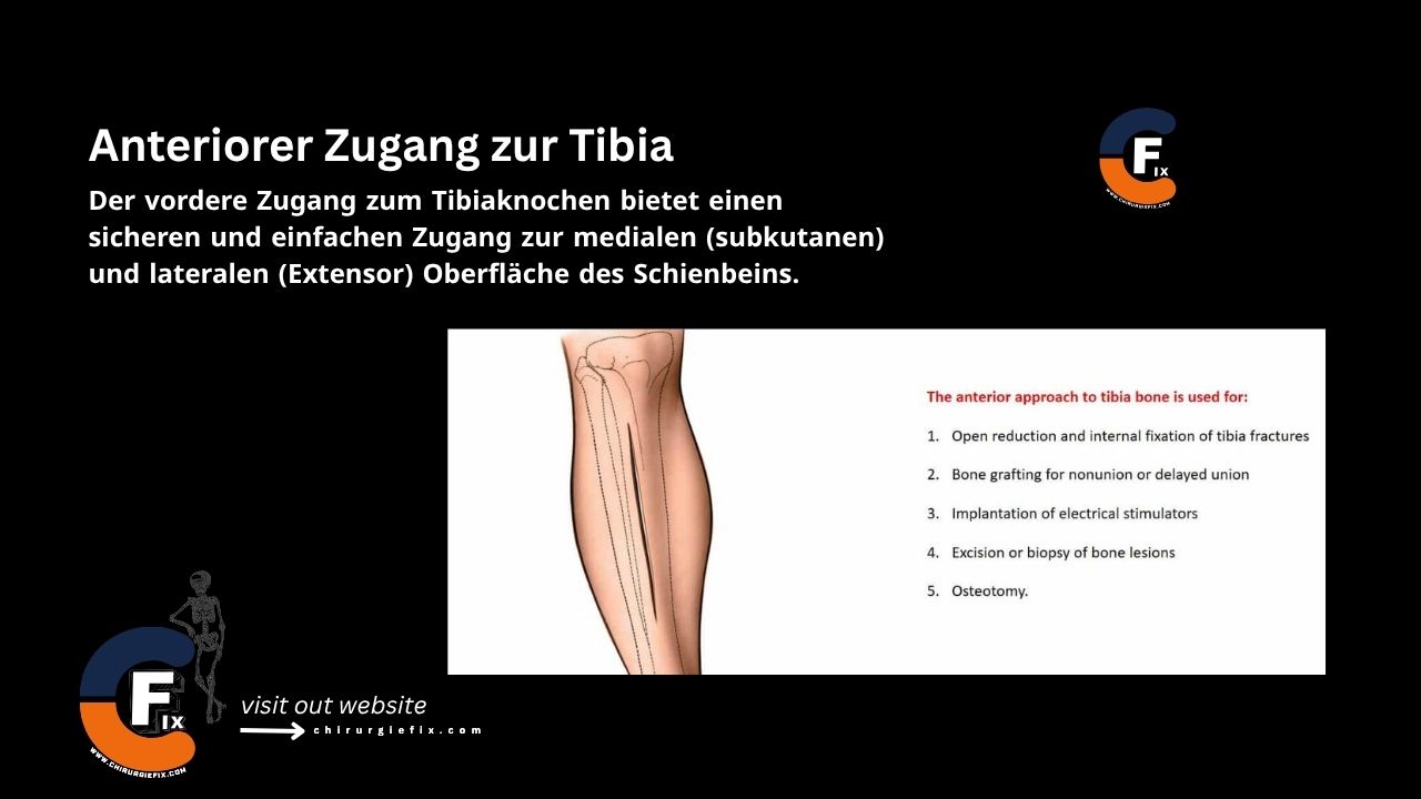
The anterior approach to the tibia provides safe and straightforward access to the medial (subcutaneous) and lateral (extensor) surfaces of the shinbone.

The anterior approach to the tibia is used for:

  • Open reduction and internal fixation of tibial fractures
  • Bone grafting for nonunion or delayed healing
  • Implantation of electrical stimulators
  • Excision or biopsy of bone lesions
  • Osteotomy

Plates applied to the subcutaneous surface of the tibia are biomechanically correctly placed on the medial (tension) side of the bone and are also easier to contour there. However, some surgeons prefer to use the lateral surface for plating to avoid the problems associated with subcutaneous placement.

The anterior approach to the tibia is the preferred approach to the shinbone unless the skin is scarred or has draining sinuses.

Patient Positioning

  • Place the patient supine on the operating table. The use of a tourniquet is optional.

Landmarks and Incision

  • Landmarks:
    1. Shaft of the tibia.
  • Incision:
    • Make a longitudinal incision on the anterior surface of the leg, parallel to the anterior border of the tibia and about 1 cm lateral to it.
    • Due to the poor blood supply of the skin, the length of the incision depends on the requirements of the procedure. It is safer to make a longer incision than to forcefully retract the skin edges to gain exposure.
    • The tibia can be exposed along its entire length.

Internervous Plane

  • There is no internervous plane for the anterior approach to the tibia.
  • The dissection is performed epiperiosteally between the tibialis anterior muscle and the tibia.

Superficial Dissection

  • Elevate the skin flaps to expose the medial (subcutaneous) border of the tibia.
  • When retracting the skin flaps, take care to protect the great saphenous vein.

Deep Dissection

  • Medial subcutaneous surface:
    • It is essential to minimize subperiosteal stripping.
    • Incise the periosteum longitudinally along the center of the medial border.
    • Reflect the periosteum anteriorly and posteriorly.
  • Lateral extensor surface:
    • Incise the periosteum over the anterior border of the tibia.
    • Subperiosteally dissect the tibialis anterior muscle and the neurovascular bundle, retracting them laterally.

Approach Extension

Proximal Extension:

  • To extend the approach proximally, continue the skin incision along the medial side of the patella. Deepen the incision through the medial patellar retinaculum to access the knee joint and the patella.
  • Alternatively, the wound can be extended proximally along the lateral side of the patella. Deepen the wound through the lateral patellar retinaculum to access the lateral compartment of the knee.

Distal Extension:

  • To extend the approach distally, make a curved incision across the medial side of the posterior aspect of the foot. Deepening the wound allows access to all structures that pass behind the medial malleolus. Continue the incision on the middle and anterior part of the foot.

Dangers

Structures at risk during the anterior approach to the tibia include:

  1. Great saphenous vein: It runs along the medial side of the leg and is vulnerable during superficial surgical dissection. It should be preserved whenever possible for future vascular procedures.

Special Surgical Points

  • Skin flaps must be carefully closed after surgery to prevent infection of the tibia. While longitudinal incisions over the shinbone heal well, transverse incisions and irregular wounds can heal poorly, especially in older patients.
  • The skin over the lower third of the tibia is very thin; wounds in this area heal poorly, especially in patients with chronic venous insufficiency.
  • It is important to minimize the amount of soft tissue stripped from the bone during the anterior approach to the tibia when it is used for fracture work.
  • Devascularized bone will not unite, no matter how well it is reduced and fixed. With care and the use of appropriate reduction clamps, it is usually possible to preserve all soft tissue attachments except on the smallest bone fragments.

References

  • Book "Surgical Exposures in Orthopaedics" – 4th Edition
  • Campbell's Book "Operative Orthopaedics", 12th Edition

Still to read...