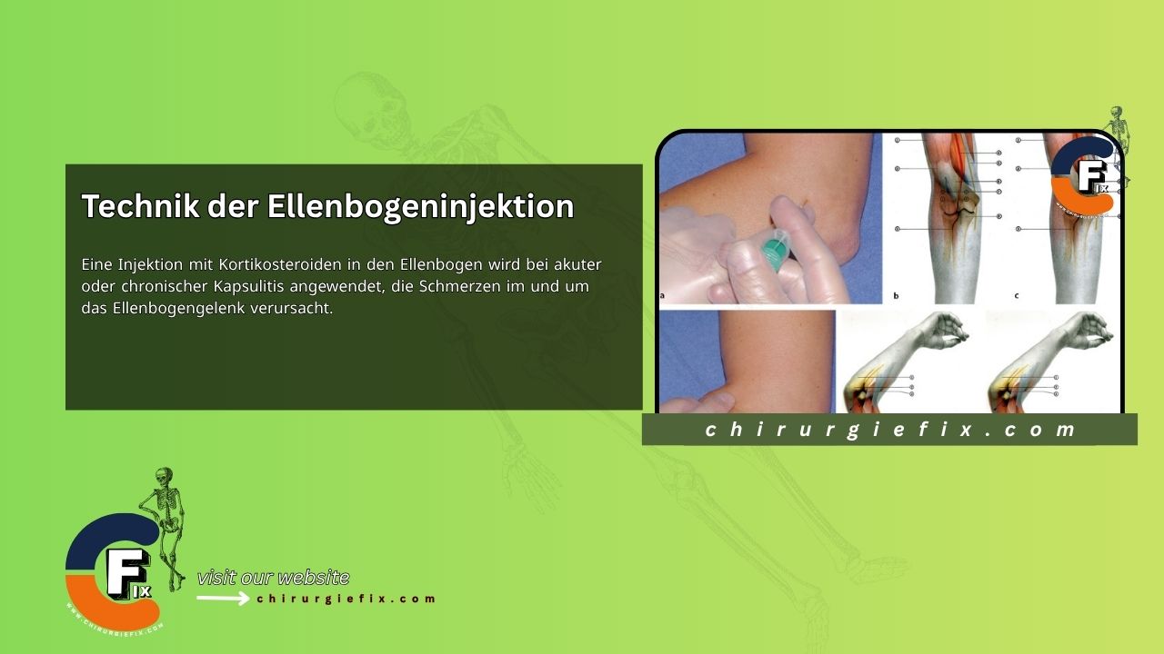
Corticosteroid injections into the elbow are used to treat acute or chronic capsulitis that causes pain in and around the elbow joint.
The capsule of the elbow joint encloses all three joints: the radiohumeral, radioulnar, and humeroulnar joints. The posterior approach, accessing the small gap between the tip of the radial head and the capitulum of the humerus, is the safest and simplest method.
To perform an elbow joint injection, follow these steps:
Begin early to increase the range of motion within pain limits using gentle stretching exercises, especially in flexion. Passive mobilization techniques are effective for achieving full range of motion but should be used with caution to avoid further joint trauma.
Elbow injections are uncommon but may be useful after trauma or a fracture of the radial head. If symptoms are caused by one or more loose bodies within the joint, the treatment involves mobilization under strong traction. If mobility improves but pain persists, an elbow injection may be considered. Adolescents with loose bodies should be referred for surgical removal.
In cases of severe joint degeneration, osteophytes at the joint margin may make needle access difficult. By injecting a small amount of the solution into the capsule, the clinician can enter the joint area with minimal discomfort for the patient. Some clinicians prefer a posterior approach, inserting the needle at the upper end of the olecranon and angling it obliquely and distally. However, this technique is somewhat more difficult to perform.
A corticosteroid injection into the biceps bursa and tendon insertion is used for chronic tendinitis or bursitis.
Although the biceps can be affected along its entire length, its attachment to the radial tuberosity on the anteromedial side of the radial shaft is particularly vulnerable. A small bursa is located at this site, which can become inflamed along with the tendon or on its own. The biceps insertion is identified by following the tendon distally from the elbow crease while the patient resists elbow flexion. The patient then relaxes the muscle, and the radial tuberosity can be palpated on the ulnar side of the radius while the forearm is passively pronated and supinated. This site is always very tender to palpation, even in a normal elbow.
The patient rests until pain-free before beginning a gradual biceps strengthening and stretching program. The cause of overuse should also be addressed.
Distinguishing between bursitis and tendinitis is often difficult. If pain is greater with passive flexion and pronation of the elbow than with resisted flexion, and if there is extreme tenderness to palpation, bursitis is more likely the cause.
If a double lesion is suspected, perform the bursal infiltration first and re-examine after one week. The tendon can then be injected if needed.