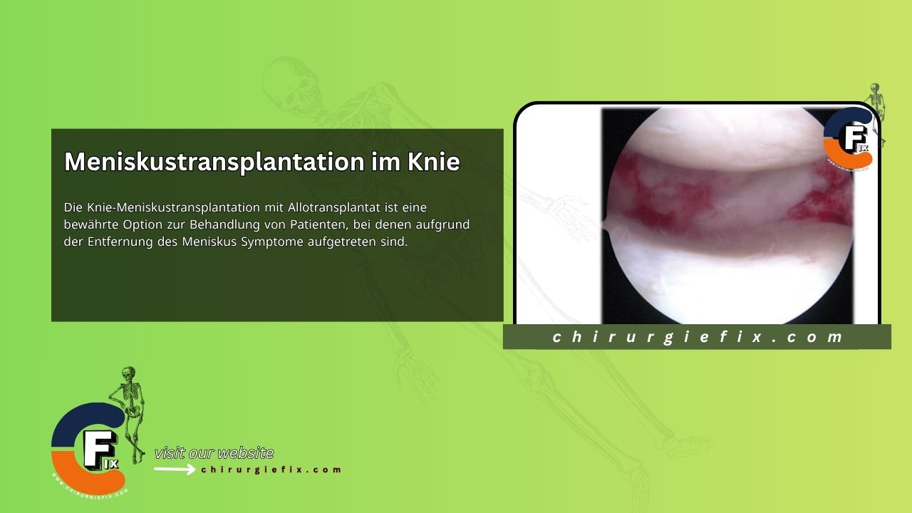
Knee meniscus transplantation using an allograft is a well-established option for patients who develop symptoms following meniscus removal. The meniscus plays a critical role in maintaining normal knee function, including shock absorption, load transmission, stability, joint lubrication, and nutrition. Consequently, partial or total meniscus removal increases the rate of degenerative arthritis in the knee. Although preserving the meniscus should be the standard of care, most tears are still treated with excision.
Since the first isolated meniscus transplantation was performed in 1984, surgical techniques have continuously improved, evolving from open procedures to arthroscopically assisted ones. While some authors have reported good results when the meniscus was fixed to the host using only soft tissue, biomechanical studies show that the allograft more closely mimics a normal meniscus when the horn attachments are secured with bone. Although it may be more technically demanding, using a bone graft-bone tunnel fixation is currently recommended. The most common techniques for achieving bony fixation of the horns are the bone bridge and bone plug methods.
A thorough patient history is essential and should include information on previous knee injuries, specific symptoms, and prior treatments or surgeries. To be considered for a meniscal allograft transplantation, the patient should have pain localized to the meniscus-deficient compartment, although joint effusions can cause general discomfort. Particular attention should also be paid to any cartilage lesions, limb alignment, and ligament stability. Attempts should be made to obtain operative reports and intraoperative photos, which can be useful in determining the amount of remaining meniscus and the condition of the articular cartilage.
Meniscus transplantation should be performed with the goal of relieving pain while increasing activity levels. Due to a lack of evidence, patients should not be told that the procedure can delay or prevent the progression of osteoarthritis.
A meniscus transplant should only be considered when non-surgical measures for pain control have been exhausted and/or there are signs that the affected compartment is degenerating. Generally, a patient should have symptoms localized to the meniscus-deficient compartment and should not have advanced arthritic changes.
Grade IV chondromalacia is a contraindication, unless it is a focal defect that can be treated with a cartilage repair or replacement method. It is generally recommended that patients be younger than 50 to 55 years; however, this is controversial, and age restrictions should also be based on the patient's physiological age.
Although these are not absolute contraindications, they must be addressed in patients with ligament laxity, limb malalignment, and full-thickness cartilage defects to be considered. The medial meniscus is known to be an important secondary stabilizer in patients with an anterior cruciate ligament (ACL)-deficient knee. Furthermore, knees with chronic ACL insufficiency have been shown to have an increased rate of meniscal tears. Therefore, in patients with ACL-deficient knees, reconstruction is required before the meniscus is replaced, or, more commonly, simultaneously with the meniscal replacement.
Limb alignment is another factor that can significantly influence the outcome of a meniscal allograft transplantation. Although there is no universal agreement, many surgeons perform an osteotomy to unload the affected compartment when the mechanical axis is several degrees greater than that of the contralateral knee. The extent of correction is typically to have the mechanical axis pass through the contralateral tibial spine, rather than the more extensive correction recommended for arthritic knees. The procedure can be performed in stages or simultaneously with the meniscus surgery, which the author prefers.
Full-thickness cartilage defects were previously considered a contraindication for meniscal allograft transplantation. However, if the defect is isolated and can be corrected through cartilage repair or restoration, it is not contraindicated.
The primary contraindication for meniscal allograft transplantation is advanced arthritis. Other contraindications include a history of infection, inflammatory arthropathy, synovial disease, and morbid obesity.
Clinical outcomes after meniscus transplantation depend on matching the size of the allograft to the natural meniscus. Oversized allografts lead to greater forces on the articular cartilage, whereas undersized allografts may result in normal forces on the cartilage but greater stress on the meniscus repair. Sizing of meniscal allografts can be performed using radiographs, computed tomography (CT), or MRI.
Sizing is most commonly based on plain radiographs using the Pollard method, which has been shown to correlate well with actual anatomical measurements.
The length of the medial and lateral meniscus is 80% and 70% of the tibial plateau, respectively. Although CT and MRI scans are more precise, they add cost with questionable benefit. A size difference of 5% is generally considered acceptable and can be achieved with proper technique and plain radiographs. Although there is no universal agreement, it is probably better for the graft to be slightly oversized, as graft shrinkage is common and has been shown to average 7%.
Four methods are available for preserving meniscal allografts: fresh, cryopreserved, fresh-frozen, and freeze-dried (lyophilized). Fresh meniscal transplants carry an increased risk of infection and immune load. Cryopreservation leads to higher costs and has not been shown to produce better results. Lyophilization alters the properties of the meniscus and is not recommended.
Therefore, fresh-frozen grafts are currently the most commonly used method for preserving meniscal allografts. It is important that allografts come from certified tissue banks, such as those accredited by the American Association of Tissue Banks, which require strict procurement and processing methods to prevent graft contamination and infection.
Further concerns have been raised regarding the age of donors for meniscal allografts, as well as the viability and tensile strength of the grafts. Current recommendations are to transplant meniscal allografts from donors younger than 45 years. It has been shown that the tensile strength of meniscal tissue, as well as its proteoglycan and collagen content, does not vary with age in grafts from donors aged 15 to 44 years.
The patient is placed supine on a standard operating table. After the induction of anesthesia, a thorough examination of the knee under anesthesia should be performed to assess range of motion and knee stability.
A tourniquet is applied to the thigh but is often not used until the arthrotomy for graft insertion. Fluid extravasation can be significant, and if the procedure is performed under dry conditions, it can lead to bleeding and obscure visualization.
Depending on the surgeon's preference, the leg can be left in a supine position without support or placed in a padded leg holder. Regardless of the technique, the posteromedial and posterolateral corners of the knee should be easily accessible for inside-out meniscal repair.
Once the patient is prepped and draped, the arthroscopic portal opposite the affected compartment should be established first. A spinal needle is used to ensure the working portal aligns directly with the horn attachments, making the tunnel easier to create straight rather than oblique. This portal will be incorporated into the arthrotomy in later stages of the procedure.
Next, a diagnostic arthroscopy is performed to assess the condition of the articular cartilage and cruciate ligaments and to confirm a meniscus deficiency. The meniscal allograft should not be opened until it is confirmed that the patient is a suitable candidate. The remaining meniscus should be trimmed back to a 1 to 2 mm peripheral rim, leaving a vascular source for healing and a reference for graft position.
If possible, the anterior and posterior horn attachments should be preserved to serve as a guide for drilling the tunnels. If visualizing the posterior attachment is difficult, a limited notchplasty should be performed.
To pass the graft, a mini-arthrotomy is made adjacent to the patellar tendon on the affected side, in line with the anterior and posterior horn insertion sites. An additional posteromedial or posterolateral incision is required for suture retrieval.
For lateral meniscus transplantation, the dovetail technique is preferred. The initial preparation is the same whether medial or lateral, and whether using a dovetail or bone plugs. Full visualization of the meniscal remnant and horn attachments is required, and a small notchplasty may be necessary to see the entire posterior root. Anatomical placement of the bone bridge is essential to prevent extrusion of the meniscal allograft.
Once the attachments are identified, a shallow (1 to 2 mm) trough is created directly in line with the anterior and posterior horns of the meniscus using a 4 mm burr. This initial trough acts as a path for placing an osteotome in the sagittal plane. A reference depth line on the osteotome helps maintain proper alignment, as the tibial plateau slopes downward when advancing from anterior to posterior. The osteotome should be advanced under direct visualization until it contacts the posterior tibial cortex.
A 6 mm drill guide is placed over the osteotome, and the drill is advanced. The guide allows the drill to be seen arthroscopically at all times, which helps maintain the correct height and prevents breaching the distal cortex. A second template is then used to accommodate a 7 mm drill bit at a height 3 mm deeper than the first template. The second drill is also visible at all times and is used to deepen the channel to 10 mm and widen the lower portion. A half-trapezoid-shaped rasp is used to complete the dovetail groove.
It is important that the rasp follows the AP slope of the tibia and that the top of the rasp remains flush with the articular surface. The channel should be cleared of any remaining bone debris, and a channel sizer should be used to ensure the preparation is complete. A posterolateral incision should then be made, utilizing the interval between the iliotibial band and the anterior edge of the biceps femoris tendon to access the capsule. While injury to the peroneal nerve should be avoided by staying anterior to the biceps femoris, the nerve can always be formally exposed.
The meniscal allograft is typically shipped from the tissue bank as a hemi-tibial plateau with the meniscus attached. It is essential to see the entire horn attachments for correct bone cuts. Therefore, excess soft tissue should be removed. The allograft bone should then be cut in an AP plane down to the ends of the attachments. Using the end of the channel rasp, an outline of the desired dovetail design is marked on the ends of the bone, serving as a reference for placing the graft in a reversed position in the holding device.
To complete the graft preparation, three cutting guides and an oscillating saw are used. The first is a vertical cut on the medial side of the graft. This is often pre-cut during meniscal allograft processing. Next, the bottom cut is aligned to match the depth of the slot. Finally, the third cutting guide is used to make the angled cut on the lateral side of the graft.
Once all cuts are complete, a sizing block is used to check that the fit is press-fit but that the graft should also slide through smoothly. Any adjustments should be made at this point, rather than attempting to force the graft into the prepared channel. To complete the graft preparation, a 2-0 polydioxanone suture is placed through the posterior segment as a reduction suture.
The reduction suture is brought out through the posterolateral incision to provide gentle traction once the graft is introduced into the tibial channel. If needed, a bone tamp can be used to gently push the bone block further into the knee until it contacts the posterior tibial wall.
At this point, the anterior arthrotomy is closed to prevent fluid leakage, and the rest of the meniscus is sutured. Various techniques can be used, but the surgeon often prefers a bucket-handle meniscal tear repair. If only an all-inside technique is used, special care must be taken to ensure the graft is not extruded outward when the fixation device is tightened. If additional fixation near the root is required, all-inside sutures are used on the most posterior side. Both absorbable and non-absorbable sutures can be used, as second-look arthroscopies have shown that graft healing is not a common problem. Typically, 8 to 10 sutures are required to fully secure the graft.
An ACL guide set to 60 degrees is used to place a guide pin into the anatomical posterior horn attachment of the old meniscus. A steep angle is chosen to assist with reducing the plug, similar to the tibial tunnel in a posterior cruciate ligament (PCL) surgery. The tibial tunnel is then drilled to a diameter of 7 mm, with the 1 mm difference allowing for easy placement of the bone plug.
A shuttle suture is passed through the tibial tunnel, retrieved, and brought out of the knee. Although the suture can be removed through the endoscope portal, the author found it simpler to perform the anterior arthrotomy before pulling the suture out. Soft tissue can often become caught between the suture and a subsequent arthrotomy incision, which can make introducing the graft into the knee difficult. A posteromedial incision is then made and dissected down to the capsule, exactly as for an inside-out medial meniscus repair.
The initial preparation of the graft is similar to the lateral side, with remaining capsular tissue attached to the meniscus removed to ensure full visibility of the horn attachments. They are then marked for size and planned orientation. Typically, an 8 mm core reamer encompasses the horn attachment. The posterior bone plug is prepared first, with a 0.062-inch K-wire inserted at approximately a 60-degree angle into the center of the attachment to mimic the angle of the tibial tunnel.
A collar pin is inserted into the pilot hole, and an 8 mm core reamer is used to create a bone plug. The end of the bone plug is then trimmed to a diameter of 6 mm and a length of 8 mm. This size has been found to adequately secure the fixation without being too large and hindering the reduction of the plug. Although a 6 mm corer can be used, the author has found it easier to use the slightly larger corer to ensure it fully encompasses the attachment, making it less likely that the plug will be damaged during harvest.
The anterior horn is harvested in a similar manner, but the plug is left at an 8 mm diameter and 10 mm length, as reduction is not difficult. To introduce and secure the posterior bone plug, a non-absorbable 2-0 suture is passed up through the bone plug, sewn through the meniscal horn attachment, and passed back down through the plug.
It is essential to incorporate the meniscal rim when suturing to give the suture additional strength, as the bone plug can be destroyed if pulled forcefully on its own. The anterior bone plug can be secured with the same suture technique or placed using a press-fit method, which will be described later. As with the dovetail technique, a polydioxanone suture is placed through the meniscus to act as a reduction suture.
The meniscal reduction sutures are introduced into the knee and retrieved through the posteromedial incision using open-ended needles and the inside-out technique. The posterior bone plug suture is passed through the tibial tunnel suture, which then guides the bone plug suture out of the tibial tunnel entrance.
The surgeon carefully places tension on the bone plug suture and gently reduces the plug with their index finger to bring the meniscus into the knee. Simultaneously, an assistant applies gentle traction to the reduction suture to guide the meniscus into the knee, which also prevents the graft from flipping over.
Although the anterior plug can be placed immediately after the posterior plug, it is advisable to first place a posterior suture, then a medial suture for initial fixation before securing the anterior horn. For example, if the anterior plug is fixed first and its placement is incorrect, a discrepancy will have to be accounted for when suturing the remaining meniscus. This can lead to an extruded graft or a graft where excessive tension is placed on the repair.
If the initial fixation is done posteriorly instead of medially, it is much easier to make adjustments for the placement of the anterior bone plug. Once the site for the anterior bone plug is determined, a guide pin is placed, and it is drilled to a depth of 1 cm with an 8 mm drill. The anterior bone plug can be secured with a suture or press-fitted.
Until experience is gained, it is advisable to use a suture, as ensuring a secure press-fit requires a learning curve. If the graft is to be secured with a bone plug suture, a small hole is drilled into the anterior tibia to accommodate a suture passer, and the plug sutures are passed through, drawing the plug into the socket. The sutures of the two plugs are then tied together for fixation. For the press-fit method, the tibial socket is made 1 to 2 mm shallower, and the plug is driven into the socket with a punch.
The suture for the posterior plug is tied at the tibial opening after reduction. The author has found that a simple, inexpensive polyethylene button works as well as a more expensive fixation device. The subsequent meniscal suturing is performed as previously described for the dovetail method.
The initial post-operative care involves controlling pain, limiting swelling, and achieving full extension. Weight-bearing is restricted for the first 4 weeks. During this time, a post-operative ROM brace is worn, with flexion limited to 90 degrees to minimize meniscal displacement and resulting stress on the posterior repair.
After 4 weeks, patients are allowed full weight-bearing and unrestricted range of motion. Low-resistance stationary cycling and swimming with straight legs are also started at this time. Once the gait pattern has normalized, progressive, gentle strengthening is performed. Most surgeons recommend waiting until 4 months before starting jogging, although the author has found no disadvantage to allowing it as early as 12 weeks. Regardless of timing, patients with moderate arthritis are strongly advised to permanently avoid strenuous activities.
From 4 to 6 months, leg strengthening is advanced to emphasize core and balance exercises. Patients are discharged after 6 months if strength and endurance are comparable to the contralateral leg and agility tests are normal. Regardless of the grade of chondromalacia, patients should not have the expectation of returning to unlimited high-performance or contact sports.