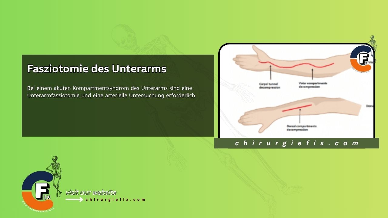
Acute compartment syndrome of the forearm requires immediate forearm fasciotomy and arterial evaluation. Compartment syndrome is a condition in which increased tissue pressure within a confined space compromises circulation and function of the structures within that space.
Forearm fasciotomy is performed using a volar approach with a curved incision technique, similar to the McConnell method for exposing the neurovascular bundles of the median and ulnar nerves. The Henry approach begins with an anterior curvilinear incision on the medial side of the biceps tendon. The incision should cross the elbow flexion crease obliquely. To allow for carpal tunnel release, extend the incision distally into the palm, but avoid crossing the wrist flexion crease perpendicularly.
Begin by dividing the lacertus fibrosus proximally and evacuating any hematomas present. If a brachial artery injury is suspected, expose the artery and assess blood flow. If flow is inadequate, remove the adventitia to reveal any underlying clots, spasm, or intimal tears. Resection of the adventitia and subsequent arterial anastomosis or grafting may be necessary.
Next, release the superficial volar compartment by using open scissors to divide the fascia covering the muscles in this compartment. Identify the flexor carpi ulnaris and retract it medially along with the underlying ulnar neurovascular bundle. Also retract the flexor digitorum superficialis and the median nerve laterally to expose the flexor digitorum profundus in its deep compartment. Check the tension of the overlying fascia or epimysium of the flexor digitorum profundus and perform a longitudinal incision if needed.
If the muscle appears gray or dark, the prognosis for recovery may be poor. Nevertheless, the muscle should still be allowed to reperfuse, as it may still be viable. Continue the dissection distally by incising the transverse carpal ligament along the ulnar border of the palmaris longus tendon and the median nerve.
In cases of median nerve palsy or paresthesia, thoroughly examine the entire zone of injury to ensure the median nerve is not severed, crushed, or trapped between the ulnar and humeral heads of the pronator teres. If such entrapment is found, perform a partial pronator tenotomy. For patients with a supracondylar fracture, reduce and stabilize the fracture with Kirschner wires and control any bleeding.
Avoid closing the skin at this stage of the forearm fasciotomy. Plan for a delayed secondary closure. If the median nerve is exposed in the distal forearm, loosely suture a distally based radial forearm flap over the nerve. Evaluate the dorsal compartments clinically or repeat pressure measurements. In most cases, volar fasciotomy alone is sufficient to decompress the dorsal musculature. However, if dorsal compartment involvement is still suspected, these compartments should also be released.
To complete the forearm fasciotomy, make an incision distal to the lateral epicondyle between the extensor digitorum communis and the extensor carpi radialis brevis. The incision should extend approximately 10 cm distally. Carefully undermine the subcutaneous tissue and release the fascia covering the mobile wad of Henry and the extensor retinaculum.
Finally, apply a sterile, moist dressing and a long arm splint, ensuring the elbow is not flexed beyond 90 degrees.