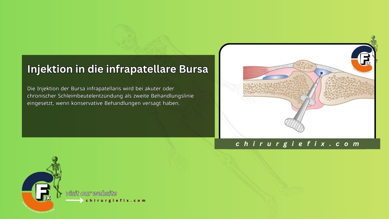
Injection into the infrapatellar bursa is used as a second-line treatment for acute or chronic bursitis when conservative treatments have failed.
Acute or chronic bursitis is caused by overuse, prolonged running or kneeling, trauma, or a direct blow to the infrapatellar bursa. It is characterized by pain in the anterior knee area distal to the lower patellar pole, painful resistance to knee extension, full passive knee flexion, and tenderness in the midline of the tendon.
There are two infrapatellar bursae: one superficial and one deep beneath the tendon. According to a limited study, the deep bursa was consistently located at the posterior side of the distal third of the tendon and was slightly wider. A fat pad apron is present, originating from the retropatellar fat pad, which partially separates the bursa. The technique described applies to the deep bursa, which is more commonly affected.
See Also: Knee Ligaments Anatomy
Begin by having the patient sit with the leg extended and the knee supported. Next, locate the tender area in the midline of the tendon and mark it. Then, insert the needle horizontally at the lateral edge of the tendon, just above the tibial tubercle. It is important to ensure the needle penetrates deeply into the posterior surface of the tendon and that there is no resistance to fluid flow. Finally, deliver the solution as a bolus.
To prevent further discomfort, it is recommended to limit weight-bearing on the knee until the pain subsides. In some cases, corrective measures such as adjusting footwear, modifying running style, and performing exercises to strengthen the quadriceps and thigh muscles may be necessary. For individuals whose professions, like carpet laying, may contribute to the problem, a cushion with a hole can be used to reduce pressure on the affected bursa.
Infrapatellar bursa injection can be performed using ultrasound guidance.
It is tempting to believe that pain in the midline of the patellar tendon is caused by tendinitis, but in our experience, this is extremely rare at this site. Infrapatellar tendinitis is regularly found at the proximal teno-osseous junction of the patella or, less commonly, at the tibial tubercle attachment. Pain here in an active adolescent should be considered Osgood-Schlatter disease and should not be injected due to the proximity to the growing end plate; taping and rest can alleviate symptoms.
For the superficial infrapatellar bursa and the prepatellar bursa, palpate the center of the tender area and inject just deep to the skin and above the bone using the same equipment. Free flow of fluid confirms correct placement within the structure. Consider using hydrocortisone in thin, dark-skinned individuals to avoid skin depigmentation or fat atrophy.