Surgery for golfer’s elbow

Understanding Golfer's Elbow Surgery

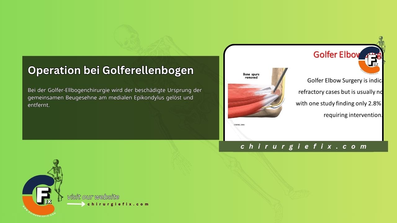
Golfer’s elbow surgery involves releasing and removing the damaged origin of the common flexor tendon at the medial epicondyle. While this procedure is indicated for refractory cases, it is not usually required. In fact, one study found that only 2.8% of patients eventually need surgical intervention.

Anatomy of the Medial Epicondyle

The medial epicondyle serves as the common origin for the flexor and pronator muscles of the forearm. The pronator teres, flexor carpi radialis, palmaris longus, and flexor digitorum superficialis all originate here and are innervated by the median nerve. The flexor carpi ulnaris also attaches at the medial epicondyle but is innervated by the ulnar nerve.

Together, these five muscles share a common origin, forming the common flexor tendon of the medial epicondyle of the humerus. This tendon is approximately three centimeters long. It crosses the medial ulnohumeral joint and runs parallel to the ulnar collateral ligament, where it acts as a secondary stabilizer.

Surgical Steps for Golfer’s Elbow

The procedure typically follows these steps:

  • Incision: Begin with a 5 cm slightly curved incision starting about 1 cm proximal and just behind the medial epicondyle. This posterior placement helps avoid the sensory branches of the medial antebrachial cutaneous nerve, which are located anterior and distal to the epicondyle.
  • Exposure: Retract the subcutaneous tissue and skin over the medial epicondyle to expose the common flexor origin.
  • Tendon Incision: To expose the pathological tissue, make a longitudinal incision in the tendon origins, starting at the tip of the medial epicondyle and extending 3 to 4 cm distally.
  • Debridement: Remove the pathological tissue elliptically, including the joint capsule if necessary. Ensure the normal tissue attachment at the medial epicondyle remains intact.
  • Closure: Close the elliptical defect with absorbable suture material.
  • Ulnar Nerve: If symptoms or pathological anatomy are identified during surgery, consider ulnar nerve transposition.
  • Wound Closure: Close the subcutaneous tissue with absorbable sutures, then close the skin with a running subcuticular suture.
  • Dressing: Apply a dressing and immobilize the elbow with a posterior splint at 90 degrees of flexion.

Postoperative Care and Rehabilitation

Recovery from golfer’s elbow surgery follows a structured timeline:

  • The splint is typically removed one week after surgery, and elbow range-of-motion exercises are started.
  • Strengthening exercises begin once full range of motion is achieved, usually around 3 weeks after the operation.
  • Strenuous activities can resume once the patient has regained normal strength without pain, which typically occurs about 3 months after surgery.
  • Patients who undergo ulnar nerve transposition usually require a longer period of immobilization and a slower progression through rehabilitation.

References and Further Reading

  1. Kiel J, Kaiser K. Golfer's Elbow. [Updated June 26, 2023]. In: StatPearls [Internet]. Treasure Island (FL): StatPearls Publishing; 2023 Jan.-. Available from: https://www.ncbi.nlm.nih.gov/books/NBK519000/
  2. Mercer's Textbook of Orthopaedics and Trauma, Tenth Edition.
  3. Sahu RL. Percutaneous release of golfer's elbow under local anaesthesia: a prospective study. Rev Bras Ortop. 2016 Sep 20;52(3):315-318. doi: 10.1016/j.rboe.2016.06.007. PMID: 28702390; PMCID: PMC5497005.

Still to read...