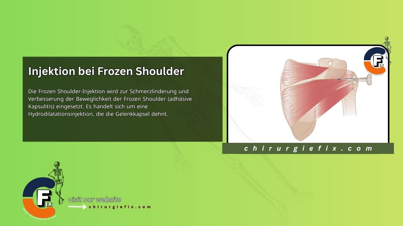
A frozen shoulder injection is used to relieve pain and improve mobility in cases of frozen shoulder (adhesive capsulitis). It is a hydrodilatation injection that stretches the joint capsule.
There are two types of frozen shoulder injection:
The shoulder joint is surrounded by a large capsule. The easiest and least painful access point is from the back, where there are no major blood vessels or nerves. An imaginary oblique line runs through the shoulder joint, from the posterior angle of the acromion forward to the coracoid process. The needle follows this line, passing through the deltoid muscle, infraspinatus, and the posterior capsule. The endpoint should be the feeling of sticky cartilage on the humeral head or glenoid.
| Syringe | Needle | Kenalog 40 | Lidocaine | Total Volume |
|---|---|---|---|---|
| 5 ml Green | 21 gauge 1.5–2 inches (40–50 mm) | 40 mg | 4 ml, 1% | 5 ml |
For larger shoulders requiring more volume, 40 mg of Adcortyl with 40 mg of 1% lidocaine can be used. Smaller patients may only need 30 mg.
After the frozen shoulder injection, maintain mobility with pendulum and stretching exercises within a pain-free range. Progress to more vigorous stretching as pain subsides. Temporary sling support and oral analgesics can be helpful in the acute stage. Once pain decreases, strong passive stretching can begin, followed by a program for rotator cuff strengthening and stabilization, along with posture correction.
Frozen shoulder typically affects patients between 40 and 60 years of age. For older patients or those with obvious signs of osteoarthritis elsewhere, an X-ray should be considered to rule out associated glenohumeral osteoarthritis (which appears normal in frozen shoulder). The less the pain radiates and the earlier the joint is treated, the more dramatic the symptom relief can be.
In the initial stage of frozen shoulder, one injection is usually sufficient, but additional injections can be safely given at increasing intervals. For advanced capsulitis, 4 to 6 injections may be administered over approximately 2 months. Inform the patient that a repeat dose may be necessary if symptoms are severe.
If resistance to the injection occurs, the needle was likely inserted too laterally and needs to be positioned more medially.
In rare cases where the posterior approach is not effective, an anterior approach can be used. The arm is held in slight lateral rotation, and the needle is inserted anteriorly between the coracoid process and the lesser tubercle of the humerus, aiming posteromedially toward the spine of the scapula with the same dose and volume. Disadvantages of this approach include the patient being able to see the needle, the skin on the flexor muscles being more sensitive, and more neurovascular structures being present on the front of the shoulder.
A range of conditions can occur alongside shoulder-neck and shoulder problems, potentially leading to diagnostic confusion. Reassessment after treating the most likely lesion may reveal another lesion. The prudent use of imaging can be helpful. There is no conclusive evidence to support or refute the use of these injections.
Two studies specifically examined steroid injection for frozen shoulder alone or compared it to a placebo injection. In a study by Roh et al., diabetic patients who received intra-articular steroid injections showed significant improvement in pain scores after four weeks compared to those who did not receive an injection (p = 0.020), although no significant difference was found after four weeks.
Significant improvements were seen in functional scores (based on ASES) (p = 0.042), forward elevation (p = 0.030), and internal rotation (p = 0.045) in those who received corticosteroid injections after 12 weeks, but not after 24 weeks. Limitations of this study included a small sample size (n = 45) and the lack of blinding of outcome assessors, which could have led to observation bias.
Three studies compared steroid injection with physiotherapy. In a study by van der Windt et al., corticosteroid injection was compared with physiotherapy, with a follow-up of 52 weeks. They did not include a placebo group. The study found significant differences at all follow-ups for all outcomes in favor of corticosteroid injection, except for the severity rating of the main complaint and abduction, where smaller differences between groups were observed at 26 and 52 weeks.
The authors also demonstrated treatment success (defined as complete recovery or strong improvement) after seven weeks in 77% of patients treated with corticosteroid injections, compared to 46% of patients treated with physiotherapy (difference between groups 31%, 95% confidence interval [CI] 14%–48%).
Only one study specifically compared intra-articular corticosteroid injection with oral NSAIDs. In this study, both the steroid injection and NSAID groups showed significant improvement in pain and range of motion (24 weeks vs. two weeks, p = 0.001). After 24 weeks, there were no significant differences between intra-articular corticosteroid injection and oral NSAIDs for all endpoints. However, this study had several limitations: unclear adequacy of allocation concealment; the lack of a placebo group for comparison; outcome assessors who were not blinded; and a high loss-to-follow-up rate (24%).
In a study by Yoon et al., intra-articular triamcinolone 40 mg (high-dose group) and intra-articular triamcinolone 20 mg (low-dose group) were compared with a placebo group. After 12 weeks, both the high- and low-dose groups showed significant improvements compared to the placebo group in pain (p < 0.001), disability (p < 0.001), and passive range of motion for flexion (p < 0.01 and p = 0.08, respectively), abduction (p < 0.001), and internal rotation (p < 0.001 and p = 0.005, respectively). After 12 weeks, there was no statistically significant difference between the high-dose and low-dose groups for all outcomes.