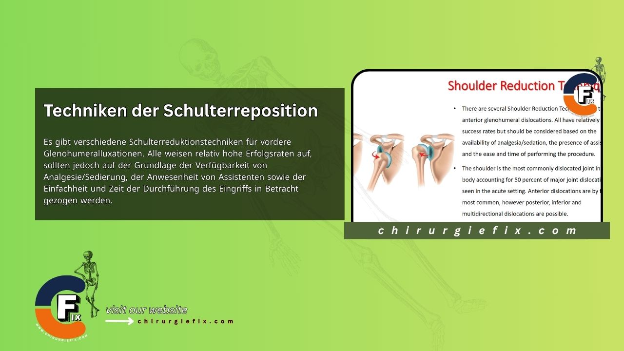
Several techniques exist for reducing anterior glenohumeral dislocations, all with relatively high success rates. The choice of technique should be guided by the availability of analgesia or sedation, the number of assistants present, and the simplicity and time required for the procedure.
The shoulder is the most frequently dislocated joint in the body, accounting for 50 percent of all major joint dislocations seen in acute care. Anterior dislocations are by far the most common, but posterior, inferior, and multidirectional dislocations can also occur.
A diagnosis of acute shoulder dislocation can be made based on the patient's history and a physical exam, though it is often confirmed with imaging. Radiographs provide a reliable assessment of the dislocation's direction. Before attempting a reduction, it is crucial to examine the injured shoulder carefully.
The axillary nerve and vascular bundle can be injured either from the initial trauma or as a complication of the reduction technique itself.
It is also essential to obtain high-quality X-rays both before and after the reduction. To confirm the presence and direction of the glenohumeral joint, an anteroposterior (AP) view, a scapular Y view, and a lateral axillary view are required.
For a primary anterior shoulder dislocation, immediate reduction will provide the patient with significant pain relief. Closed reduction can be performed with or without anesthesia. Reduction without anesthesia works best for recurrent or very recent dislocations where there is limited rotator cuff muscle spasm. Intra-articular lidocaine injection has been shown to be as effective as sedation for shoulder reduction, while limiting potential drug complications and reducing time to discharge.
Common reduction techniques for anterior dislocation include:
The patient lies prone with the affected arm hanging off the table. A 5 to 10-pound weight is attached to a strap around the patient's wrist.
The physician uses their hands to push the inferior tip of the scapula medially while moving the superior aspect laterally.
This technique is often preferred because it can successfully reduce dislocations with little or no analgesia. It can be performed with the patient lying supine or sitting upright.
When the patient is upright, the ipsilateral elbow should be supported to avoid any traction. The elbow is flexed to 90 degrees, and the arm is gently rotated externally. The physician should rotate slowly, pausing if the patient experiences pain to allow for muscle relaxation. If the shoulder has not reduced spontaneously by 90 degrees of external rotation, the arm is slowly abducted, and the humeral head may be lifted into place.
The success of this method relies on complete muscle relaxation. The patient is placed prone on a stretcher or exam table. The injured arm is allowed to hang over the side, and a 10 to 15-pound weight is attached, similar to the method described above. Reduction typically occurs within 20 to 30 minutes.
With the patient lying supine, the physician externally rotates and abducts the patient's arm. As the arm reaches an overhead position, the elbow is extended. The physician applies gentle traction. Using their free hand, the physician can manipulate the humeral head over the glenoid labrum.
The patient is placed supine. A sheet is tied around the patient's chest and positioned at the level of the axilla. An assistant provides countertraction using the sheet while the physician applies traction to the patient's forearm at an abduction angle of 30 degrees and forward flexion of 20 to 30 degrees.
Traction should be gentle and may require continuous application for up to 5 minutes. Sudden, forceful movements should be avoided, as they can cause additional neurovascular, soft tissue, or bone injury to the patient.
The patient is placed supine with the elbow flexed to 90 degrees and the arm abducted. A sheet is tied around the patient's chest and the waist of an assistant. A second sheet is placed around the patient's proximal forearm and the physician's waist.
The physician applies traction to the patient's arm while the assistant provides countertraction. Reduction using this technique may require gentle internal and external rotation or manipulation of the proximal humerus.
If the techniques described above are unsuccessful, reduction under anesthesia may be necessary. This technique may also be required when significant fractures are present. Anesthesia allows for complete muscle relaxation, and the reduction is often accomplished easily with a low risk of additional injury.
Posterior shoulder dislocations are much rarer, accounting for only 1 to 2 percent of all glenohumeral dislocations. They are often associated with a history of direct trauma to the front of the shoulder, strong muscle contractions during seizures or electric shock, or falls onto an outstretched hand.
They can be more difficult to identify on physical exam than anterior dislocations, which makes confirmation with a scapular Y view X-ray very important.
Associated bone lesions include:
Posterior dislocations are often accompanied by severe pain and muscle spasm, making analgesia and muscle relaxation extremely important.
The reduction technique for a posterior dislocation is performed with the help of an assistant. The patient lies supine. The physician applies traction to the humerus with the arm adducted. The assistant gently manipulates the humeral head forward into the glenoid.
Luxatio erecta, also known as inferior dislocation, is extremely rare. Severe soft tissue trauma and fractures commonly accompany an inferior dislocation due to the mechanism of injury.
The patient history typically describes the arm being forced into hyperabduction, with the humeral neck levered against the acromion. The acromion acts as a fulcrum, driving the humeral head downward and tearing the inferior capsule.
The physical exam is characteristic of luxatio erecta, with the arm held in full abduction. The patient's hand usually rests on or behind their head. The humeral head is often palpable on the lateral chest wall.
For this type of dislocation, reduction is achieved with an assistant using traction and countertraction. The physician applies traction in line with the humerus while the assistant provides countertraction. An audible "clunk" indicates successful reduction.
A two-step technique can also be used for reducing an inferior dislocation. In this approach, the physician first converts the luxatio erecta into an anterior dislocation, after which a preferred technique for anterior reduction is used to complete the procedure.
The patient is placed supine. Sedation can be administered if needed. The physician stands next to the patient's head on the ipsilateral side of the injury. Facing the patient's feet, the physician places the hand closer to the patient (upper hand) on the midshaft of the humerus, while the opposite hand (lower hand) is positioned over the medial epicondyle.
The lower hand applies a gentle upward force on the distal humerus while the physician uses the upper hand to manipulate the humeral head from its inferior position toward the anterior rim of the glenoid. A straight shoulder contour and a prominent posterolateral edge of the acromion indicate that the humeral head has been moved anteriorly.
At this point, the physician should be able to adduct the humerus. The physician can then use whichever anterior reduction technique they find most comfortable.
After subluxations and dislocations, short-term shoulder immobilization should be recommended based on the patient's pain level.
For primary dislocations, an early program focusing on range of motion and rotator cuff strengthening should be recommended. However, extreme external rotation or forward flexion should be avoided.
A decrease in the fear of external rotation and abduction is often a good indicator that a patient may be able to return to normal activities once their strength has also improved.
Patients under 20 years of age are very likely to develop recurrent dislocations due to soft tissue injuries sustained during their first episode. The rate of recurrent instability is often considered inversely proportional to age.
Early arthroscopic Bankart repair has been proposed for primary anterior dislocations, showing positive results in the young, active patient population, with fewer recurrences of instability.