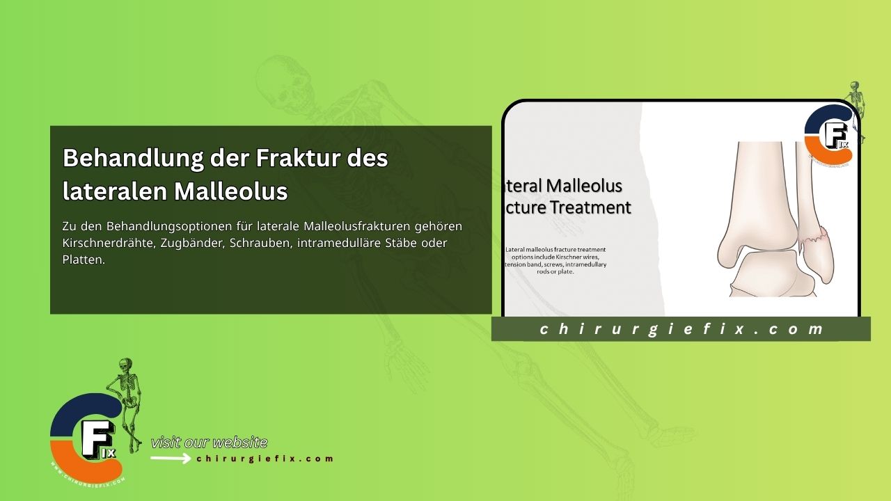
Treatment options for lateral malleolus fractures include Kirschner wires, tension bands, screws, intramedullary rods, or plates.
When the broken fibula is part of a bimalleolar fracture pattern, the lateral malleolar or fibular fracture is typically reduced and internally fixed before the medial malleolar component is fixed. An exception is when the lateral malleolus is comminuted as part of a bimalleolar or trimalleolar pattern. In severe cases of comminution, excessive reduction of the lateral malleolus can occur, which hinders the anatomical reduction of the medial malleolar component. In such cases, it may be advisable to proceed with medial malleolar fixation first.
To expose the lateral malleolus and the distal fibular shaft, make a lateral longitudinal incision, taking care to protect the superficial peroneal nerve. Alternatively, a posterolateral incision can be made, and the plate can be inserted using a posterior antiglide technique.
If the fracture is sufficiently oblique, has good bone stock, and no comminution, fix the fracture with two lag screws inserted from anterior to posterior for interfragmentary compression. Place the screws approximately 1 cm apart. Screw length is critical to ensure engagement of the posterior cortex for secure fixation without protruding far enough posteriorly to interfere with the peroneal tendon sheaths.
For transverse fractures, an intramedullary device can be used. Split the fibers of the calcaneofibular ligament longitudinally to expose the tip of the lateral malleolus. Insert a Rush rod, an interlocking fibular rod, or another intramedullary device across the fracture line into the medullary canal of the proximal fragment. Avoid tilting the lateral malleolus toward the talus, as this can cause narrowing of the ankle mortise and limited range of motion. Contour the intramedullary nail to prevent this error.
When the fracture is below the plafond level, the distal fragment is small, and the patient has good bone stock, use an intramedullary 3.5 mm malleolar screw for fixation. In larger patients, a 4.5 mm lag screw can be used. Alternatively, you can angle the malleolar screw slightly obliquely to engage the medial cortex of the fibula proximal to the fracture.
In patients with poor bone quality, Kirschner wires can be placed obliquely from lateral to medial through the distal and proximal fibular fragments. These wires can be further secured with a tension band wire.
Anatomical reduction and restoration of fibular length are crucial aspects of the procedure.
For fractures above the level of the syndesmosis, after achieving anatomical reduction, use a small fragment, one-third tubular plate for fixation. For larger individuals or more proximal fractures, a 3.5 mm dynamic compression plate can be used. Plates can supplement fixation with lag screws or span a comminuted segment. Generally, three cortical screws are placed in the fibular shaft above the fracture and two or three screws distal to the fracture. Unicortical cancellous screws are placed below the plafond. With posterolateral placement, the plate acts as an antiglide plate. Although commercially available pre-contoured distal fibular locking plates with fixed angles offer alternative fixation options in the distal area, they often lead to increased hardware prominence.
In cases involving osteoporotic patients or those with poor soft tissue coverage, reduce and stabilize the fracture with Kirschner wires inserted obliquely through the distal fibular fragment and into the tibia. Syndesmotic fixation.
For immobilization of the ankle after lateral malleolus fracture surgery, a posterior splint is applied in a neutral position and elevated.
If bone quality is satisfactory and fixation is secure, the splint can be replaced with a removable brace or fracture boot at the first postoperative visit. Range of motion exercises are initiated while weight-bearing is limited for 6 weeks. After this time, if fracture healing is progressing properly, partial weight-bearing can begin and advance accordingly.
However, if skin conditions, bone quality, or other factors prevent secure fixation, the fracture requires extended protection. In such cases, the patient is placed in either a short-leg or long-leg, non-weight-bearing cast, depending on the stability of the fixation. If a long-leg cast is used initially, it can be changed to a short-leg cast after 4 to 6 weeks. Weight-bearing on the ankle is strictly prohibited until sufficient fracture healing has occurred (8 to 12 weeks). At this stage, a short-leg walking cast is worn, and weight-bearing is gradually increased. The cast is removed once the fracture is fully united.