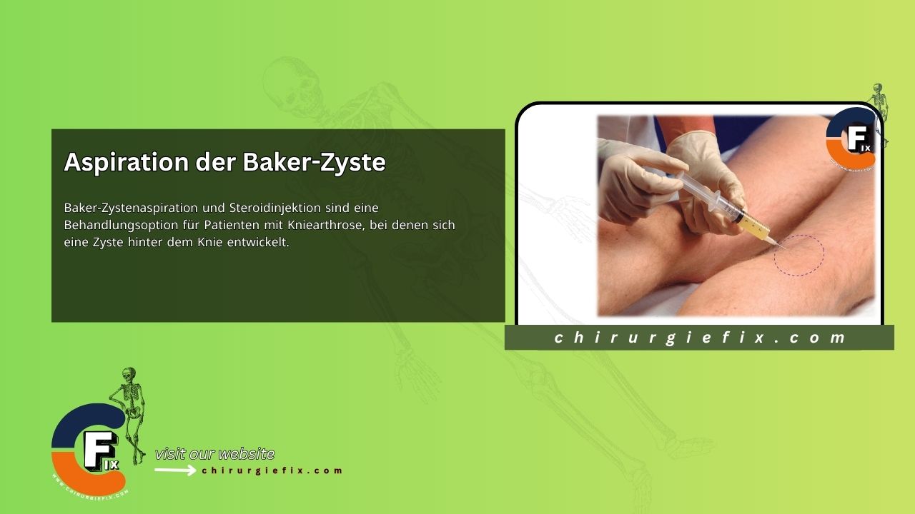
Baker’s cyst aspiration combined with a steroid injection is a treatment option for patients with knee osteoarthritis who develop a cyst behind the knee. This procedure can help relieve discomfort and improve mobility.
A Baker’s cyst appears as a noticeable, often large swelling in the popliteal fossa (the hollow at the back of the knee). It can limit both active and passive knee bending.
These cysts are most common in adults aged 35 to 70 and are frequently linked to inflammatory joint conditions such as rheumatoid arthritis or osteoarthritis. They can also result from overuse or a knee injury. Because many Baker’s cysts are asymptomatic, they are often discovered incidentally during a physical exam or imaging, such as an MRI performed to investigate suspected osteoarthritis or other internal knee issues. The prevalence of Baker’s cysts typically increases with age, likely due to a greater communication between the knee joint and the bursa that develops over time.
A Baker’s cyst is an enlarged sac of synovial fluid. It forms when fluid pushes through a defect in the posterior wall of the knee joint capsule or results from an effusion within the semimembranosus bursa. The popliteal artery and vein, along with the posterior tibial nerve, run centrally through the popliteal fossa. These structures must be carefully avoided during the injection.
After the aspiration, a firm compression bandage may be applied for 1 to 2 days.
If the aspirated fluid is anything other than clear synovial fluid, a sample should be sent for culture and appropriate treatment should be initiated. The swelling often returns at some point, but it can be re-treated if the patient desires.
A Baker’s cyst is often found alongside a knee effusion and may disappear when the knee is aspirated, indicating that the two compartments communicate. Do not insert a needle into a pulsating Baker’s cyst, as this almost certainly indicates a popliteal artery aneurysm.
A ruptured Baker’s cyst can mimic the symptoms of a deep vein thrombosis in the calf.