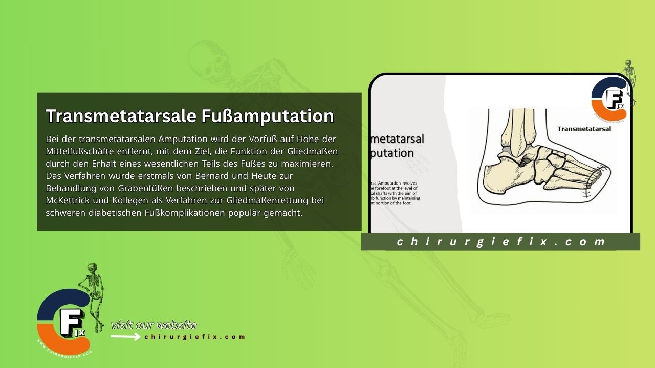
Transmetatarsal amputation (TMA) involves removing the forefoot at the level of the metatarsal shafts. The goal is to maximize limb function by preserving a substantial portion of the foot. This procedure was first described by Bernard and Heute for treating trench foot, and later popularized by McKettrick and colleagues as a limb-salvage technique for severe diabetic foot complications.
TMA is generally preferred over hindfoot amputation or traditional below-knee amputation (BKA). It is widely regarded as an effective salvage procedure for forefoot infections, gangrene, and chronic ulcers.
Creating the skin flaps: To form long plantar and short dorsal full-thickness flaps, begin the dorsal incision at the level of the intended bone cut on the anteromedial side of the foot. Curve it slightly distal to the bone cut level, reaching the midpoint of the lateral side of the foot.
Plantar incision: Start the plantar incision at the same point as the dorsal incision. Carry it distally beyond the metatarsal heads, then curve it proximally to end at the midpoint of the lateral side of the foot. The incision on the medial side should be slightly longer than on the lateral side, as the larger cross-sectional diameter medially requires more skin coverage. Design the plantar flap to include subcutaneous fat and a layer of plantar muscles.
Removing the toes and metatarsals: Disarticulate the toes at the metatarsophalangeal joints. Cut the metatarsal bones obliquely from dorsal-distal to plantar-proximal at the junction of the middle and distal thirds.
Cascading the metatarsals: The metatarsal bones should be resected in a cascading manner. The second metatarsal osteotomy should be only a few millimeters shorter than the first. Each subsequent cut should be 2 to 3 mm shorter than the previous medial metatarsal. The fifth metatarsal should be shorter still—4 to 5 mm shorter than the fourth. Always use a power saw to resect the metatarsal bone to prevent later bone overgrowth. Use a rongeur and a rasp to smooth any bone prominences. If infection is present distally, try to avoid disrupting any abscess and keep the metatarsophalangeal joint intact.
Managing nerves and tendons: Identify the nerves and divide them well proximally so that their cut ends sit proximal to the bone ends. Divide the tendons under tension so they retract into the foot. Alternatively, you can suture the flexor and extensor tendons together to create a myoplasty. Use a drain if necessary.
Closing the wound: Bring the long plantar flap over the bone ends and suture it to the dorsal flap with interrupted, non-absorbable sutures. Be cautious when “contouring” skin tags at the medial and lateral edges, as this can compromise the blood supply to the flap. This excess tissue will resolve over time.
Dressing and immobilization: Apply a light compression dressing and place the foot in a carefully padded posterior splint, with the ankle in neutral to slight dorsiflexion.