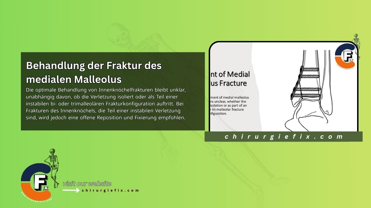
The optimal treatment for fractures of the medial malleolus remains a subject of debate, whether the injury occurs in isolation or as part of an unstable bi- or trimalleolar fracture pattern. However, open reduction and internal fixation is recommended for medial malleolus fractures that are part of an unstable injury.
Begin the incision approximately 2 cm proximal to the fracture line. Extend it distally and slightly posteriorly, ending about 2 cm distal to the tip of the medial malleolus.
This incision is preferred in the surgical treatment of medial malleolus fractures for two reasons:
Note: Distal extension of the incision may not be possible if concomitant foot injuries need to be addressed.
Handle the skin gently, ensuring that the flap and underlying subcutaneous tissue remain intact. Because blood supply to this skin area is poor, careful handling is required to prevent skin sloughing. Also protect the great saphenous vein and its associated nerve.
The distal fragment of the medial malleolus is usually displaced distally and anteriorly, often with a small periosteal fold lying between the fracture surfaces. Remove this fold from the fracture site using a curette or a periosteal elevator, exposing the small fracture spikes.
Remove small, loose bone or cartilage fragments. Preserve and support large osteochondral fragments with a bone graft.
Use a bone-holding clamp or a towel clamp to bring the displaced malleolus into its normal position. While holding it in place, perform internal fixation using two smooth 2-mm Kirschner wires, drilled across the fracture site as temporary fixation devices.
Check fracture reduction using anteroposterior and lateral radiographs. If the reduction is satisfactory, proceed as follows:
Alternatively, a drill with 2.5-mm and 3.5-mm bits can be used to create a path for the screws. A long pelvic drill may be required for bicortical lag screw fixation. Carefully inspect the interior of the joint, particularly the superomedial corner, to ensure the screw has not penetrated the articular surface. Verify screw and fracture position with radiographs.
If the medial malleolar fragment is very small or comminuted, screw fixation may not be possible. In such cases, use multiple Kirschner wires or tension band wires for fixation. For large vertical fractures with proximal comminution, a buttress plate may be necessary to prevent loss of reduction. A small, one-third tubular plate is usually sufficient. Exercise extreme caution when applying bulky hardware to avoid wound complications in areas with poor skin coverage.
For immobilization of the ankle after medial malleolus fracture fixation, apply a posterior plaster splint in a neutral position and elevate the limb. If bone quality is satisfactory and fixation is secure, the splint can be replaced with a removable splint or fracture boot at the first postoperative visit. Range-of-motion exercises are initiated while weight-bearing is limited for 6 weeks. After this period, partial weight-bearing can begin when fracture healing is adequate and can progress accordingly.
However, if skin conditions, bone quality, or other factors prevent secure fixation, the fracture requires extended protection. In such cases, the patient is fitted with either a short-leg or long-leg, non-weight-bearing cast, depending on the stability of the fixation. If a long-leg cast is initially used, it can be changed to a short-leg cast after 4 to 6 weeks. Weight-bearing on the ankle is strictly prohibited until the fracture has progressed sufficiently to heal (8 to 12 weeks). At this stage, a short-leg walking cast is worn, and weight-bearing is gradually increased. The cast is removed once the fracture is fully united.