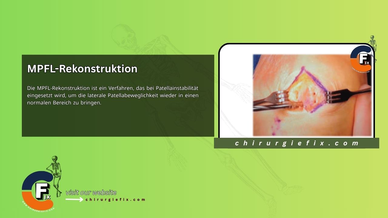
MPFL reconstruction is a surgical procedure used to treat patellar instability by restoring lateral patellar mobility to a normal range. The medial patellofemoral ligament (MPFL) is the primary ligamentous restraint against lateral patellar displacement, providing an estimated 50% to 60% of the medial soft tissue resistance to lateral patellar dislocation. Consequently, a patellar dislocation often injures the medial retinacular structures, including the MPFL, leading to increased lateral patellar mobility. A functional MPFL is both necessary and sufficient to bring lateral patellar mobility back into a normal range; therefore, surgical treatment should focus on restoring a functional MPFL.
Indications for MPFL Reconstruction
Non-operative treatment is typically indicated for acute, first-time dislocations without an associated osteochondral fracture or loose body. Surgical treatment is indicated for the following:
- Recurrent patellar instability (at least 2 documented instability events) with a confirmatory physical exam showing excessive lateral patellar laxity: For these recurrent dislocators, MPFL reconstruction is the current procedure of choice.
- Osteochondral fracture or loose body that is potentially repairable or causing mechanical symptoms: Fragment repair must consider the size and location of the donor fragment and the presence of sufficient subchondral bone. When a surgically treated osteochondral injury is present, a concurrent MPFL reconstruction may be performed.
- Small, isolated cartilage injuries: Surgery may be indicated if mechanical symptoms are present. If no mechanical symptoms are present, non-operative management is appropriate.
MPFL Reconstruction Procedure
Key Imaging Considerations for Preoperative Planning
Patellar Height
- The preferred measurement method is that of Caton and Deschamps (the ratio between the distance from the inferior edge of the patellar articular surface to the superior edge of the tibial plateau and the length of the patellar articular surface).
- A ratio of 1.2 or greater indicates patella alta, which predisposes a patient to patellar instability because the patella engages the trochlea late in knee flexion.
- If patella alta is present, tibial tubercle osteotomy and distalization should be considered as an additional procedure.
Tibial Tubercle–Trochlear Groove (TT-TG) Distance
- The TT-TG offset is the transverse distance between the tibial tuberosity and the center of the trochlear groove.
- This distance can be measured on axial CT or MRI, although some argue these imaging modalities are not interchangeable as MRI has been reported to underestimate the TT-TG offset compared to CT.
- If the lateral offset is 20 mm or greater, medialization of the tibial tubercle may be considered as an additional procedure to treat patellofemoral instability.
Trochlear Morphology
- An important consideration, as severe cases of trochlear dysplasia may compromise the long-term success of MPFL repair.
- Can be assessed on a true lateral radiograph (where the posterior borders of both femoral condyles are strictly superimposed).
- A crossing sign occurs when the floor of the trochlea crosses the anterior border of both femoral condyles, indicating trochlear dysplasia.
- A positive trochlear prominence (the sagittal distance between the trochlear groove and the anterior femoral cortex) of more than 3 mm on the lateral view has been shown to correlate well with trochlear dysplasia.
- Axial MRI has been shown to classify trochlear groove anatomy more accurately in the assessment of trochlear dysplasia (compared to axial radiographs or CT).
Equipment
For the authors' preferred method of MPFL reconstruction, the following equipment is recommended in addition to standard soft tissue retractors:
- Knee arthroscopy equipment, if knee arthroscopy is to be performed prior to MPFL reconstruction
- Soft tissue tendon stripper for harvesting the semitendinosus graft (the authors prefer a closed tendon stripper)
- 4.5 mm drill with guide for drilling tunnels in the patella for each end of the semitendinosus tendon graft
- Small curved curette to clear the patellar tunnels of debris for graft passage
- 18-gauge steel wire shaped to pull suture through the patellar tunnels. Commercially available curved suture-passing devices are alternative options.
- 2.4 mm diameter smooth guidewire pin (Beath pin equivalent), with an eyelet for suture if needed. This pin is used to drill into the center of the femoral MPFL footprint and guide the reamer for creating the femoral MPFL tunnel. If the pin is too long, it may interfere with fluoroscopy to confirm anatomical positioning of the pin at the MPFL origin.
- Fluoroscopy to verify the position of the guide pin in the femoral footprint of the MPFL. The authors prefer using a mini C-arm equivalent.
- Cannulated drills of various diameters from 5 to 8 mm for drilling the femoral tunnel
- Interference screws for graft fixation in the femoral tunnel. Typically, screws with lengths of 20 to 25 mm and diameters of 5 to 8 mm are used.
- Resorbable suture material No. 0 or No. 1
- #5 braided polyethylene suture material
- No. 2 non-resorbable suture material
Positioning and Portals
The patient is positioned supine. A small bump may be placed under the hip to prevent external rotation. A sterile bump can also be placed under the knee to maintain slight flexion. If knee arthroscopy is performed before MPFL reconstruction, it is recommended to do so with the bed flat so the knee can be flexed over the edge of the bed, rather than lowering the foot of the bed for knee flexion.
Arthroscopy can be performed to diagnose or treat joint cartilage lesions and to rule out pre-existing severe arthritis if there is doubt about the cartilage health of the patellofemoral joint. If needed, fluoroscopy can be performed from the ipsilateral side of the bed to obtain a lateral view of the distal femur and confirm the anatomical placement of the guide pin in the femoral MPFL footprint.
Description of the Procedure
Examination Under Anesthesia
Perform an examination under anesthesia to confirm excessive lateral patellar mobility. Increased laxity is indicated by lateral translation greater than 10 mm and the absence of a firm endpoint with the knee flexed at 30 degrees.
Harvesting and Preparing the Semitendinosus Tendon Graft
- The sartorial fascia is exposed through a 2 to 3 cm skin incision made 2 cm medial and distal to the medial border of the tibial tubercle.
- The sartorial fascia is incised along the palpable gracilis tendon. Avoid cutting too deeply to prevent injury to the underlying superficial medial collateral ligament.
- Identify and isolate the gracilis tendon (proximal) and the semitendinosus tendon (distal) from their deep side (the bursal layer).
- Apply tension to the semitendinosus while releasing it from the crural fascia at the posteromedial corner using tissue scissors.
- Place holding sutures of resorbable No. 0 or No. 1 suture on a tapered needle, then transect the tendon from its tibial attachment.
- Once all tendon slips are freed, the semitendinosus tendon is harvested with a closed (preferred) or open tendon stripper.
- Baseball stitches using No. 2 non-resorbable suture are placed on both free ends to allow the graft to be passed through the two patellar tunnels later.
- The graft is prepared on the back table by first sizing the graft to 240 mm and then folding it in half, resulting in a doubled graft of 120 mm. It is recommended to size the doubled graft length to at least 120 mm to ensure sufficient graft length.
- A No. 5 polyethylene suture is placed through the loop to pull the doubled graft into the blind femoral tunnel.
- A 25 mm long baseball stitch with resorbable No. 0 or No. 1 suture is placed into the looped end of the graft to sew together the two graft strands that will sit in the femoral tunnel.
Patellar Tunnel Placement
- A longitudinal incision is made over the length of the patella at the junction of the medial and middle thirds of the patella (in line with the medial border of the patellar tendon at the distal patellar pole).
- The medial 8 to 10 mm of the patella is exposed through subperiosteal dissection using a No. 15 scalpel.
- The dissection extends medially and dorsally around the patella through layers 1 (longitudinal retinaculum) and 2 (native MPFL), ending after the transverse fibers of the native MPFL are cut. The capsule (layer 3) remains intact.
- A 4.5 mm drill hole is made on the medial side of the superior pole of the patella adjacent to the articular margin.
- A corresponding drill hole is made on the anterior surface of the patella approximately 8 mm from the medial border. (This point corresponds to the lateral edge of the original retinacular dissection.)
- The two drill holes are connected using a curved curette.
- A second 4.5 mm drill hole is made on the medial side of the patella at a point measuring two-thirds of the patellar length.
- A corresponding drill hole is made on the anterior surface of the patella approximately 8 mm from the medial border, and the two holes are connected with a curved curette.
- If the semitendinosus graft has a diameter greater than 4.5 mm, the drill holes are slightly enlarged to facilitate graft passage.
- It is important not to place the distal patellar tunnel distal to the natural insertion of the MPFL to avoid restricting the distal pole of the patella.
Femoral Tunnel Placement and Isometry Check
- A skin incision is made just anterior to the palpable ridge connecting the medial femoral epicondyle and the adductor tubercle.
- The knee is slightly flexed to facilitate palpation of this landmark (flexion moves the hamstring muscles posteriorly away from the medial epicondyle).
- If the landmarks are difficult to palpate, a small skin incision is made and palpation is performed through the wound to identify the ridge.
- The graft can be placed between layers 1 and 2 or between layers 2 and 3 (joint capsule) (i.e., it can lie superficial or deep to the native MPFL).
- Placement of the graft between layers 2 and 3 is preferred because blind dissection superficial to the native MPFL can disrupt the insertion of the vastus medialis obliquus into the anterior part of the MPFL; furthermore, by placing the graft deep to the native MPFL, the native MPFL can be repaired over the graft during wound closure.
- The graft should not be placed deep to the capsule, as it should remain extra-articular to avoid graft abrasion and allow complete healing.
- Using a long, curved clamp, the chosen interval (preferably between layers 2 and 3) is developed from the patellar incision anteriorly to the medial femoral epicondyle posteriorly.
- With the tip of the clamp positioned over the ridge between the medial epicondyle and the adductor tubercle, layers 1 and 2 are incised with a No. 15 blade.
- The tip of a Beath pin is placed at a point approximately 9 mm proximal and 5 mm posterior to the medial epicondyle; the pin is then driven to the lateral side of the femur. The accuracy of pin placement should be confirmed with lateral fluoroscopic images, with the ideal tunnel position being approximately 1 mm anterior to the extension line of the posterior cortex, 2.5 mm distal to the posterior origin of the medial femoral condyle, and proximal to the level of the posterior point of the Blumensaat line on a lateral radiograph with both posterior condyles projected in the same plane.
- A loop of No. 5 braided polyethylene suture is passed around the Beath pin, through the prepared retinacular tunnel, and then through one of the patellar tunnels.
- The knee is taken through its range of motion (ROM) to assess isometry while holding the suture at the anterior side of the patellar tunnel to evaluate changes in suture length during ROM.
- If lengthening occurs in flexion, a second Beath pin is placed more distally toward the medial epicondyle. The first pin is left in place to facilitate repositioning while drilling the second Beath pin. The loop of No. 5 suture is passed around the second Beath pin, and the knee is again taken through ROM. If isometry is acceptable, the first Beath pin is removed.
- If lengthening occurs in extension, a second Beath pin is placed more proximally toward the adductor tubercle. The first pin is left in place to facilitate repositioning while drilling the second Beath pin. The loop of No. 5 suture is passed around the second Beath pin, and the knee is again taken through ROM. If isometry is acceptable, the first Beath pin is removed.
- Once the femoral pin site is accepted, a blind tunnel the size of the doubled graft is drilled into the femur. For a semitendinosus graft, this is typically 6 to 7 mm in diameter.
- The femur is reamed to a depth of at least 20 mm, preferably 25 mm.
Graft Passage and Fixation
- The No. 5 suture placed through the graft loop is passed through the eyelet of the Beath pin, and the pin is then advanced out of the lateral femoral cortex to pull the graft into the femoral tunnel.
- Secure fixation to the femur can be achieved using a 20–25 mm interference screw.
- The looped isometry suture, if left in the retinacular tunnel, can be used to guide the free ends of the graft through the previously created retinacular space.
- The free graft arms are individually passed through their respective patellar tunnels using a double 18-gauge stainless steel wire or a curved suture-passing instrument.
- The graft arms enter the medial border of the patella and exit anteriorly to be overlapped for suture fixation.
- The free graft arms are folded back and sutured together just medial to the patella with 2 figure-of-eight mattress sutures using No. 2 non-resorbable suture on a tapered needle.
- After the first suture is placed, patellar mobility is checked. There should be a good endpoint or checkrein with the knee in full extension and at 30 degrees of flexion, full knee range of motion, and 7 to 9 mm of lateral patellar translation from the centered position at 30 degrees of flexion.
- Excess graft is sharply excised.
- The native MPFL is sutured to the graft, and then the retinaculum is closed over the graft.
- The wounds are closed in the usual manner.
Postoperative Protocol
For recovery following MPFL surgery, these post-MPFL reconstruction protocols should be followed:
- Immediately postoperatively, weight bearing as tolerated is allowed in a drop-lock or knee immobilizer brace.
- Passive ROM and closed-chain strengthening exercises are started as soon as possible to restore ROM and quadriceps control. Maintaining motion is critical after MPFL surgery.
- Bracing may be continued while walking for up to 6 weeks to prevent falls until quadriceps control is restored. Measuring the quadriceps circumference 10 cm above the superior pole of the patella can be helpful in assessing quadriceps recovery.
- Patients are allowed to return to stressful activities, including sports, when they have achieved full range of motion and regained at least 80% of their quadriceps strength compared to the uninjured extremity.
- If at least 90 degrees of flexion is not achieved within 6 postoperative weeks, the intensity of the therapy program must be increased. Manipulation under anesthesia may be required between 9 and 12 weeks post-surgery if stiffness cannot be resolved with therapy alone.
References
- Conlan T, Garth WP Jr, Lemons JE. Evaluation of the medial soft-tissue restraints of the extensor mechanism of the knee. J Bone Joint Surg Am. 1993 May;75(5):682-93. doi: 10.2106/00004623-199305000-00007. PMID: 8501083.
- Panagiotopoulos E, Strzelczyk P, Herrmann M, Scuderi G. Cadaveric study on static medial patellar stabilizers: The dynamizing role of the vastus medialis obliquus on the medial patellofemoral ligament. Knee Surg Sports Traumatol Arthrosc. 2006;14(1):7-12.
- Hautamaa PV, Fithian DC, Kaufman KR, Daniel DM, Pohlmeyer AM. Medial soft tissue restraints in lateral patellar instability and repair. Clin Orthop Rel Res. 1998;(349):174-182.
- Fithian DC, Gupta N. Patellar instability: Principles of soft tissue repair and reconstruction. Tech Knee Surg. 2006;5:19-26.
- Mäenpää H, Lehto MU. Patellar dislocation. The long-term results of nonoperative management in 100 patients. Am J Sports Med. 1997;25(2):213-217.
- Nikku R, Nietosvaara Y, Aalto K, Kallio PE. Operative treatment of primary patellar dislocation does not improve medium-term outcome: A 7-year follow-up report and risk analysis of 127 randomized patients. Acta Orthop. 2005;76(5):699-704.
- Nikku R, Nietosvaara Y, Kallio PE, Aalto K, Michelsson JE. Operative versus closed treatment of primary patellar dislocation: Similar 2-year results in 125 randomized patients. Acta Orthop Scand. 1997;68(5):419-423.
- Fithian DC, Paxton EW, Stone ML, et al. Epidemiology and natural history of acute patellar dislocation. Am J Sports Med. 2004;32(5):1114-1121.
- Teitge RA, Faerber WW, Des Madryl P, Matelic TM. Stress radiographs of the patellofemoral joint. J Bone Joint Surg Am. 1996;78(2):193-203.
- Panagiotopoulos E, Strzelczyk P, Herrmann M, Scuderi G. Cadaveric study on static medial patellar stabilizers: The dynamizing role of the vastus medialis obliquus on the medial patellofemoral ligament. Knee Surg Sports Traumatol Arthrosc. 2006;14(1):7-12.
- Elias DA, White LM, Fithian DC. Acute lateral patellar dislocation at MR imaging: Injury patterns of medial patellar soft-tissue restraints and osteochondral injuries of the inferomedial patella. Radiology. 2002;225(3):736-743.
- Caton J, Deschamps G, Chambat P, Lerat JL, Dejour H. Patella infera. A propos de 128 cas [in French]. Rev Chir Orthop Reparatrice Appar Mot. 1982;68(5):317-325.
- Dejour H, Walch G, Nove-Josserand L, Guier C. Factors of patellar instability: An anatomic radiographic study. Knee Surg Sports Traumatol Arthrosc. 1994;2(1):19-26.
- Camp CL, Stuart MJ, Krych AJ, et al. CT and MRI measurements of tibial tubercle–trochlear groove distances are not equivalent in patients with patellar instability. Am J Sports Med. 2013;41(8):1835-1840.
- Salzmann GM, Weber TS, Spang JT, Imhoff AB, Schöttle PB. Comparison of native axial radiographs with axial MR imaging for determination of trochlear morphology in patients with trochlear dysplasia. Arch Orthop Trauma Surg. 2010;130(3):335-340.
- Schöttle PB, Schmeling A, Rosenstiel N, Weiler A. Radiographic landmarks for femoral tunnel placement in medial patellofemoral ligament reconstruction. Am J Sports Med. 2007;35(5):801-804.
- Elias JJ, Cosgarea AJ. Technical errors during medial patellofemoral ligament reconstruction could overload medial patellofemoral cartilage: A computational analysis. Am J Sports Med. 2006;34(9):1478-1485.
- Muneta T, Sekiya I, Tsuchiya M, Shinomiya K. A technique for reconstruction of the medial patellofemoral ligament. Clin Orthop Rel Res. 1999;(359):151-155.
- Nomura E, Horiuchi Y, Kihara M. Medial patellofemoral ligament restraint in lateral patellar translation and reconstruction. Knee. 2000;7(2):121-127.